
In the second lecture of our Pachychoroid Spectrum series, we focus on the modern classification of Central Serous Chorioretinopathy (CSC) and the multimodal imaging findings that have significantly improved our understanding of this disease.
The lecture begins with the recently proposed classification system for CSC, highlighting the transition from the traditional “acute” and “chronic” terminology to the contemporary concepts of simple and complex CSC. The rationale behind this classification is discussed in detail, emphasizing why disease duration alone is insufficient and why multimodal imaging findings provide a more accurate reflection of disease severity, prognosis, and treatment planning.
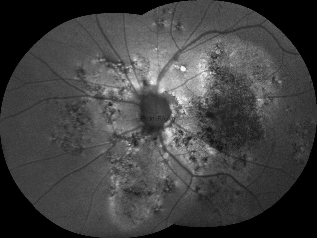
The imaging section reviews the characteristic findings of CSC using multimodal imaging. Optical coherence tomography (OCT), fundus autofluorescence (FAF), fluorescein angiography (FA), indocyanine green angiography (ICGA), and OCT angiography (OCTA) are discussed with a practical, clinically oriented approach. Particular attention is given to the interpretation of pigment epithelial detachments, flat irregular PEDs, fibrin, photoreceptor alterations, and autofluorescence patterns that distinguish acute from chronic disease. The role of OCT angiography in detecting silent macular neovascularization, which may remain undetectable with conventional angiography, is also reviewed.
The lecture also introduces several recently described imaging biomarkers and anatomical concepts that have reshaped our understanding of pachychoroid diseases. Wide-field OCT findings, asymmetric vortex vein drainage, intervortex venous anastomoses, choroidal flow void analysis, and the concept of vortex vein overload choroidopathy are discussed together with emerging evidence regarding scleral thickness, choroidal fluid loculation, and ciliochoroidal effusion. These recent discoveries provide new insights into the underlying pathophysiology of CSC and help explain why the disease extends far beyond the macular region.
This lecture provides both a practical guide for clinicians and an up-to-date review of the evolving concepts in CSC classification and imaging, serving as the foundation for the subsequent lecture on evidence-based treatment strategies.
Credit: M. Giray Ersoz, MD, FEBO, Retina Specialist
Memorial Bahçelievler Hospital, Department of Ophthalmology, Istanbul, Turkey
Arel University School of Medicine, Department of Ophthalmology, Istanbul, Turkey
Instagram accounts: @retina.review and @retina.dr.girayersoz
Website: www.girayersoz.com.tr
