
A 29-year-old man was referred to our retina clinic for photodynamic therapy (PDT) with a presumed diagnosis of central serous chorioretinopathy (CSC).
Fifteen months earlier, he had undergone pneumatic retinopexy for a rhegmatogenous retinal detachment at another institution. According to the available medical records, the retinal break was successfully sealed and the retinal tear was anatomically reattached following pneumatic retinopexy and subsequent laser retinopexy. However, a localized pocket of subretinal fluid persisted at the macula.
OCT imaging obtained two weeks after surgery already demonstrated persistent, although decreasing, subretinal fluid beneath the fovea. The amount of fluid gradually decreased over time, but never completely resolved. The patient was subsequently evaluated elsewhere, where the persistent subfoveal fluid was interpreted as chronic CSC, and he was referred to our clinic for verteporfin PDT.
At presentation, the patient’s best-corrected visual acuity was 20/22.
Multimodal Imaging
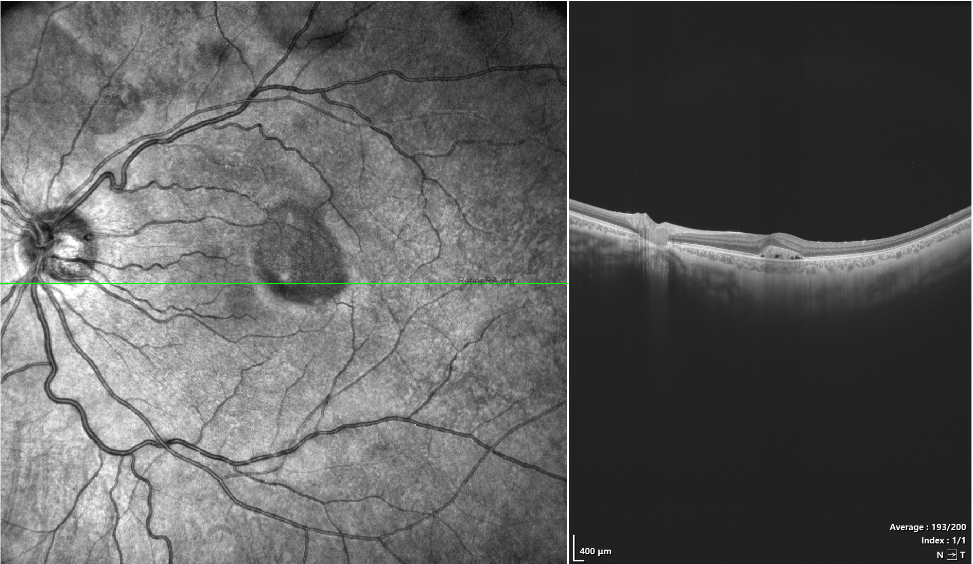
Spectral-domain OCT demonstrated a small, shallow pocket of subfoveal subretinal fluid confined to the fovea, accompanied by elongation of the photoreceptor outer segments, a morphology that closely resembles chronic CSC.
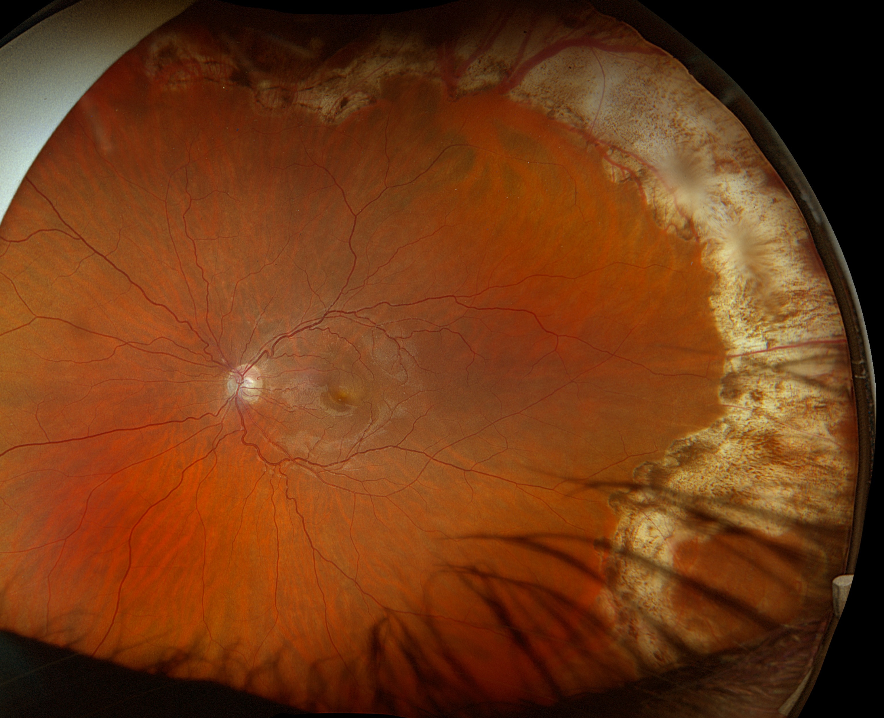
Ultra-widefield color fundus photography demonstrated extensive 360-degree laser photocoagulation scars, consistent with previous retinal detachment repair.
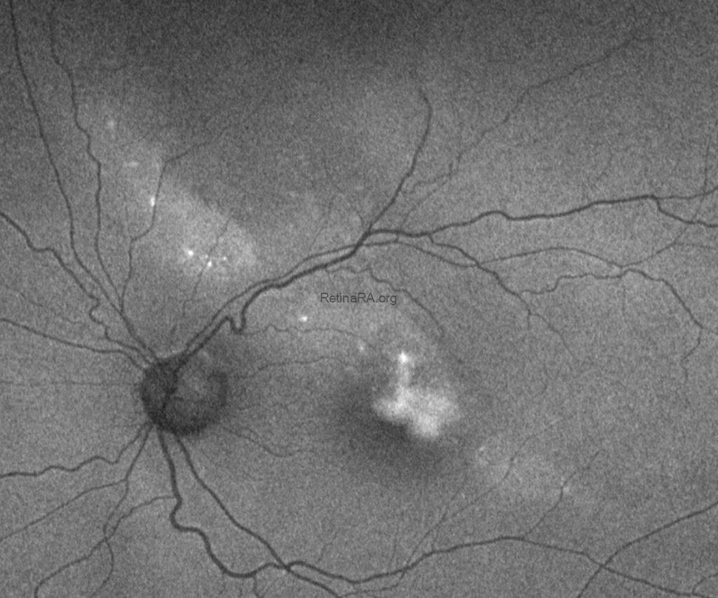
Fundus autofluorescence (FAF), however, revealed a remarkable finding. A long, narrow hyperautofluorescent tract originated approximately 4–5 disc diameters superior to the optic disc and extended obliquely toward the macula. The lesion measured approximately one disc diameter in width and seven disc diameters in length.
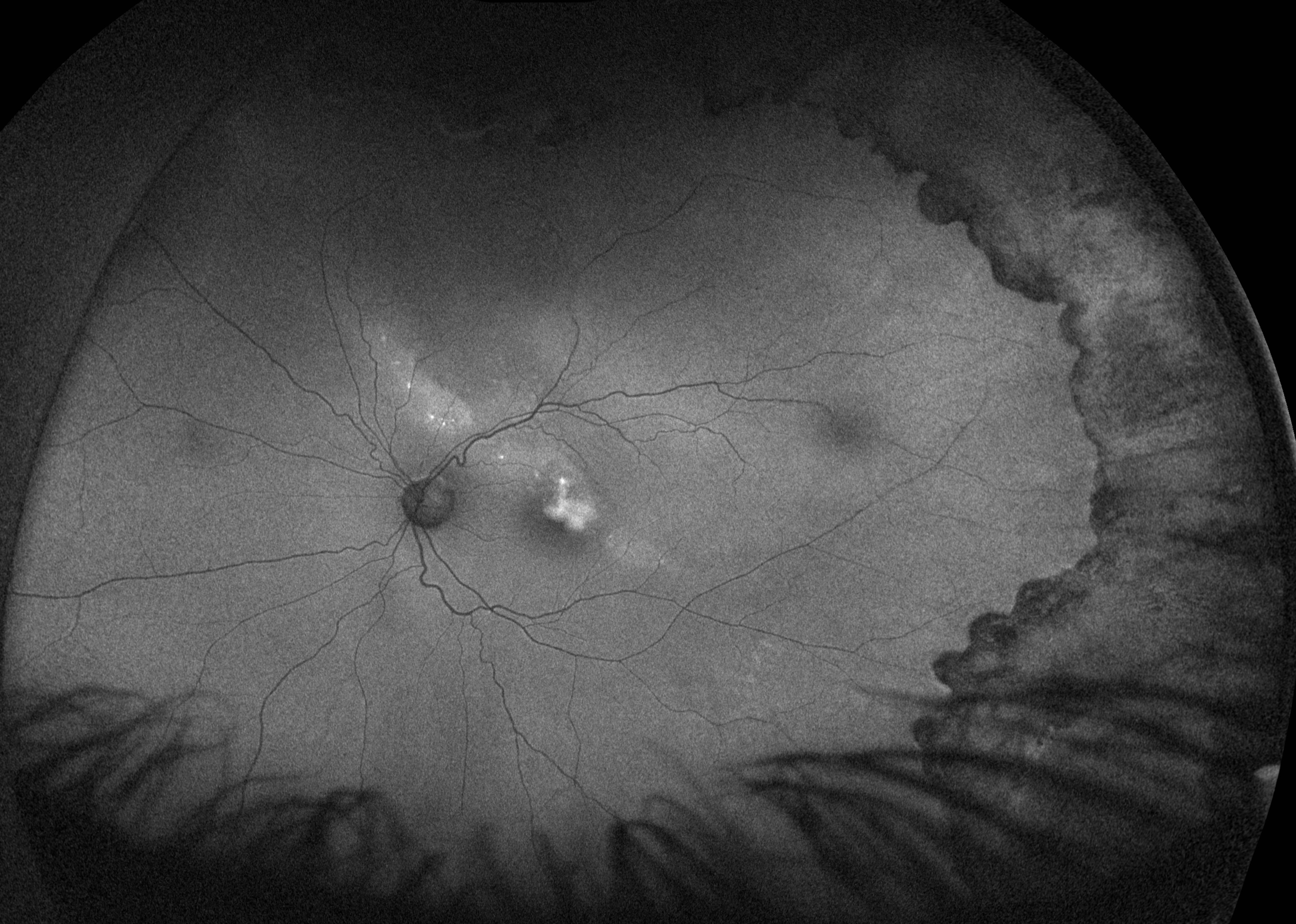
Importantly, this autofluorescence pattern was not compatible with the classic gravitational descending tracts typically observed in chronic CSC, suggesting that another diagnosis should be considered.
The Diagnostic Clue
A detailed review of the patient’s previous imaging proved decisive.
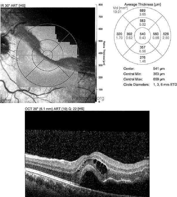
When the OCT obtained two weeks after pneumatic retinopexy was compared with the current multimodal imaging, the area of postoperative residual subretinal fluid corresponded precisely to the hyperautofluorescent tract observed on FAF.
Preoperative OCT Scan
Postoperative OCT Scan (15th day after the surgery)
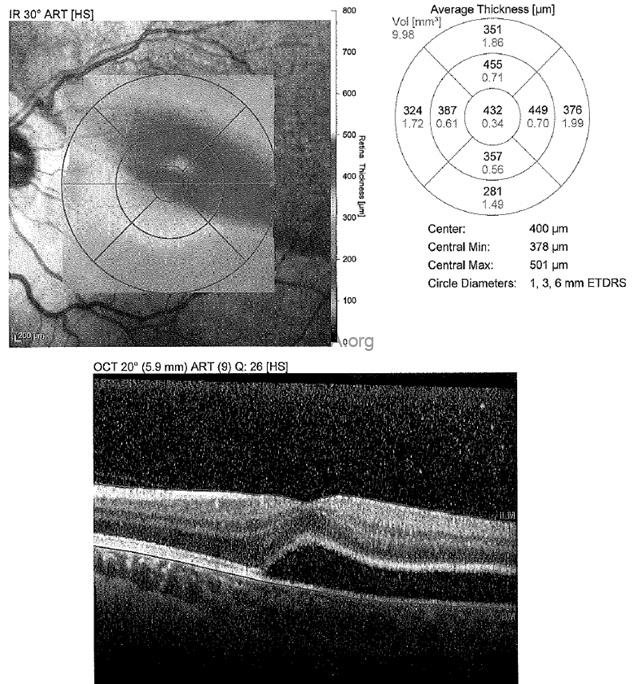
This spatial correlation strongly suggested that the shallow subfoveal fluid seen at presentation was not the result of a newly developed CSC, but rather the remaining portion of the original postoperative residual subretinal fluid, which had gradually regressed over the preceding 15 months.
Further questioning revealed that the patient had not been fully compliant with the recommended postoperative positioning following pneumatic retinopexy, a factor that may have contributed to the prolonged persistence and distribution of the residual subretinal fluid.
The diagnosis was established not by a single image, but by comparing multimodal images obtained 15 months apart.
Final Diagnosis
The patient was diagnosed with persistent residual subretinal fluid following successful pneumatic retinopexy, rather than central serous chorioretinopathy.
Because the imaging findings were entirely explained by the previous retinal detachment repair, photodynamic therapy was not performed, and careful observation was recommended.
Discussion
Residual subretinal fluid is a well-recognized finding following successful repair of rhegmatogenous retinal detachment. Although spontaneous absorption occurs in most eyes, complete resolution may require several months and, in some patients, may persist for more than one year. Younger patients and eyes with macula-off retinal detachments appear particularly prone to prolonged residual subretinal fluid.
Pneumatic retinopexy remains an excellent treatment option for appropriately selected rhegmatogenous retinal detachments, particularly those with superior retinal breaks. The procedure offers excellent anatomical outcomes while avoiding more invasive surgery. Nevertheless, meticulous postoperative positioning remains a critical component of successful treatment and may influence both anatomical recovery and the rate of subretinal fluid absorption.
Persistent subfoveal fluid after retinal detachment repair may closely mimic chronic CSC because both conditions can demonstrate shallow subretinal fluid and elongation of the photoreceptor outer segments on OCT. However, careful evaluation of the patient’s surgical history, serial OCT examinations, retinal laser scars, and fundus autofluorescence characteristics usually allows the correct diagnosis to be established.
In the present case, the hyperautofluorescent tract did not exhibit the morphology of a gravitational tract typical of chronic CSC. Instead, it precisely matched the distribution of the postoperative residual subretinal fluid documented shortly after retinal detachment repair, providing the key diagnostic clue.
Multimodal imaging is powerful, but longitudinal multimodal imaging is often decisive.
Recognizing this entity is clinically important because it can prevent unnecessary interventions such as verteporfin photodynamic therapy.
Teaching Points
- Persistent residual subretinal fluid following pneumatic retinopexy may remain detectable for many months and occasionally for longer than one year.
- OCT findings alone may closely resemble chronic central serous chorioretinopathy.
- Hyperautofluorescent patterns that do not follow the typical gravitational configuration of CSC should prompt reconsideration of the diagnosis.
- Comparing current images with early postoperative OCT scans may reveal the true origin of persistent subretinal fluid.
- A detailed surgical history combined with longitudinal multimodal imaging can prevent unnecessary treatments such as photodynamic therapy.
Take-home message: Not every pocket of subfoveal fluid is central serous chorioretinopathy. Sometimes, the correct diagnosis lies not in the current OCT, but in the patient’s previous images.
Credit: M. Giray Ersoz, MD, FEBO, Retina Specialist
Memorial Bahçelievler Hospital, Department of Ophthalmology, Istanbul, Turkey
Arel University School of Medicine, Department of Ophthalmology, Istanbul, Turkey
Instagram accounts: @retina.review and @retina.dr.girayersoz
Website: www.girayersoz.com.tr
