
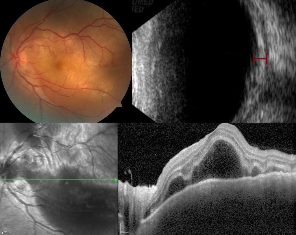
A 30-year-old man presented with pain and blurred vision in his left eye. Best-corrected visual acuity was 1.0 in the right eye and 0.3 in the left eye, with intraocular pressures within normal limits. There were no inflammatory cells in the anterior chamber or vitreous.
Fundus examination revealed optic disc edema, choroidal folds, and diffuse macular elevation with whitish-yellow discoloration at the posterior pole in the left eye; while the right eye was completely normal.
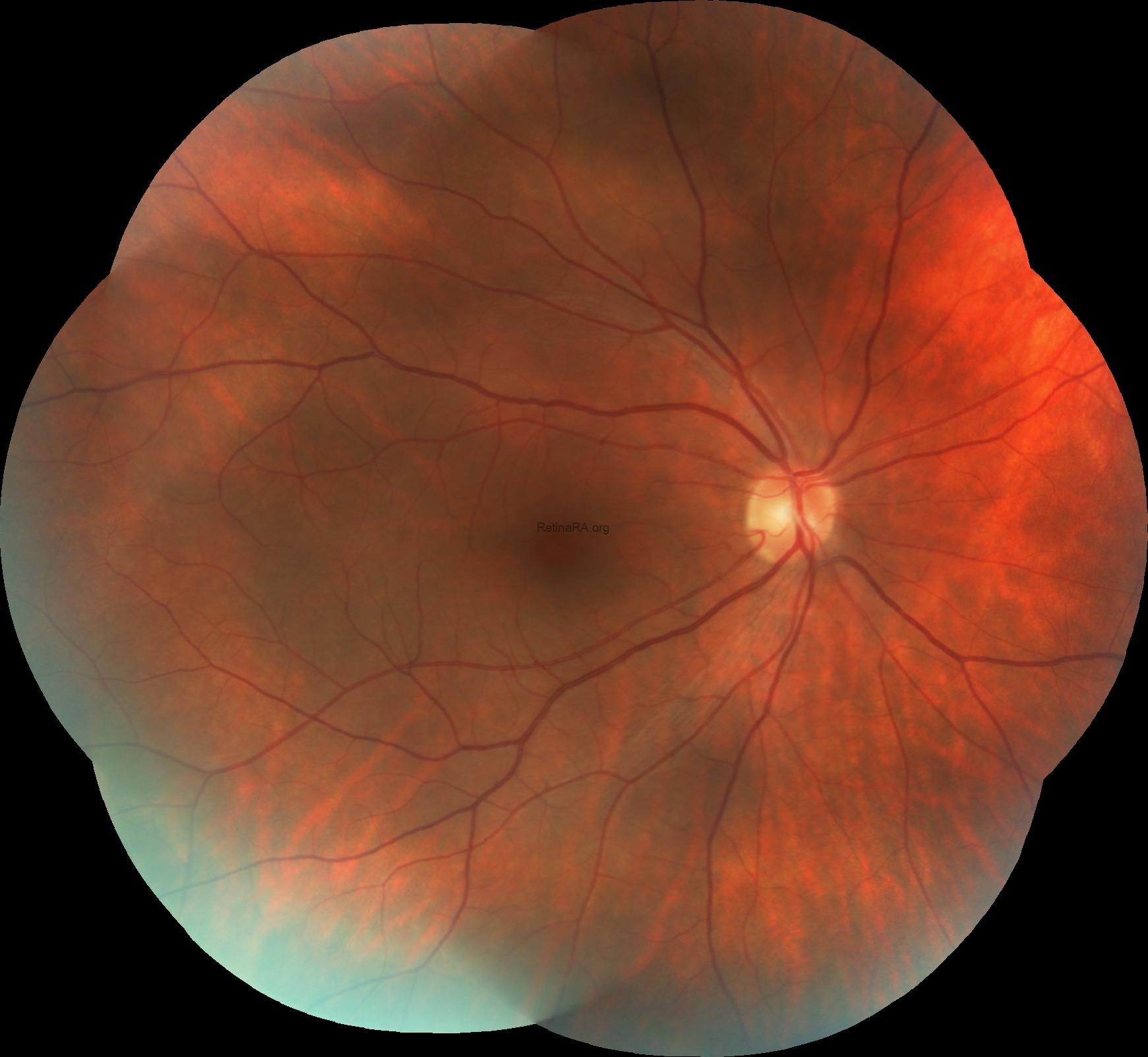
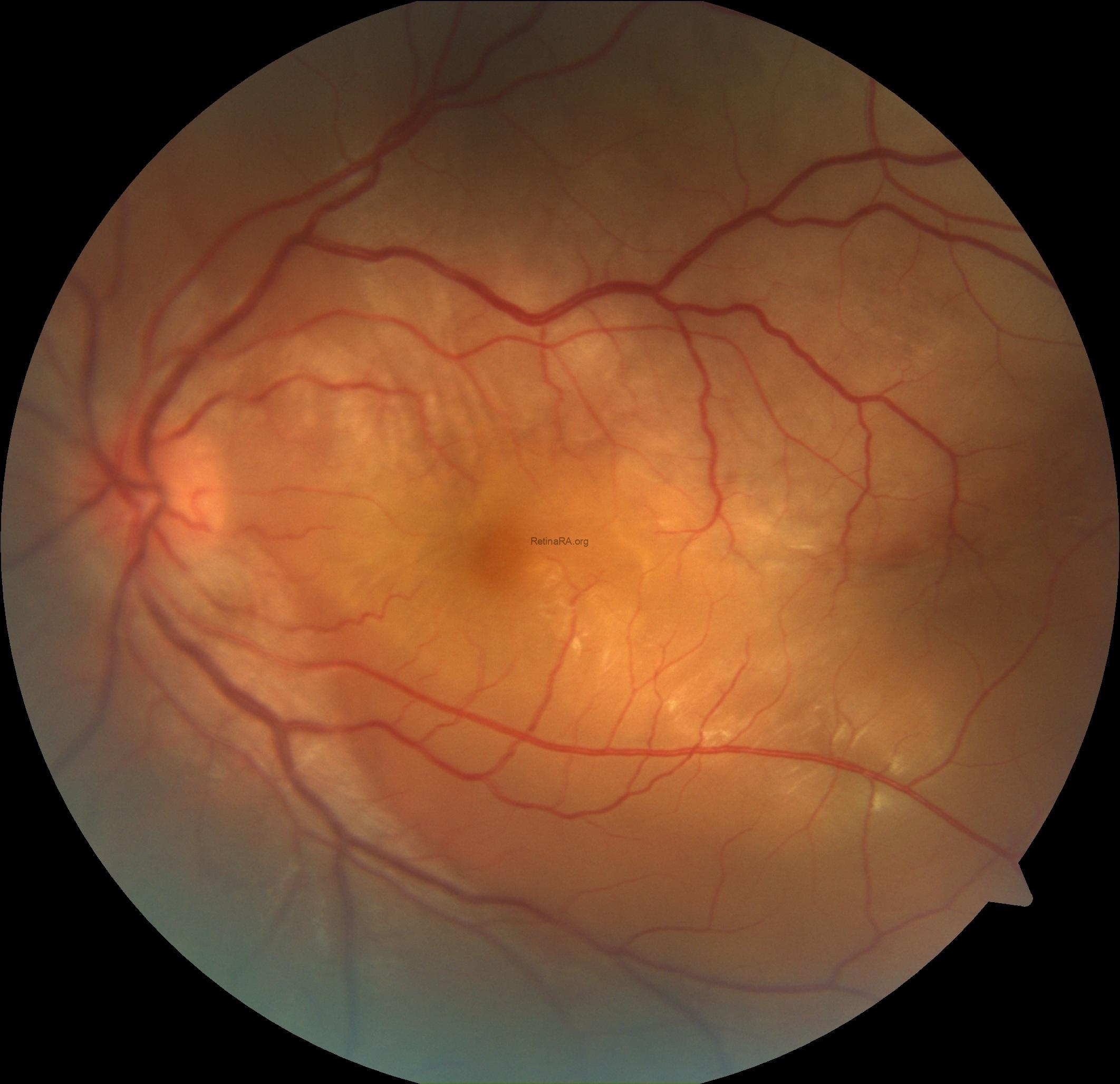
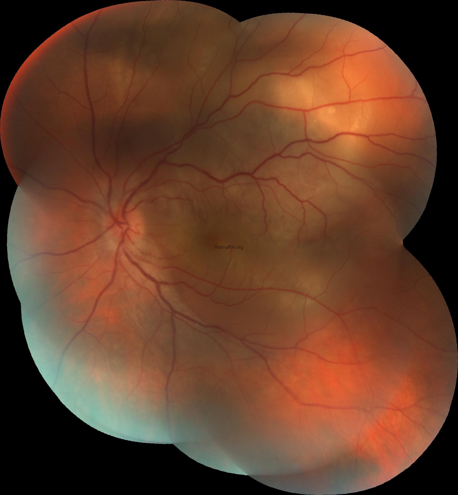
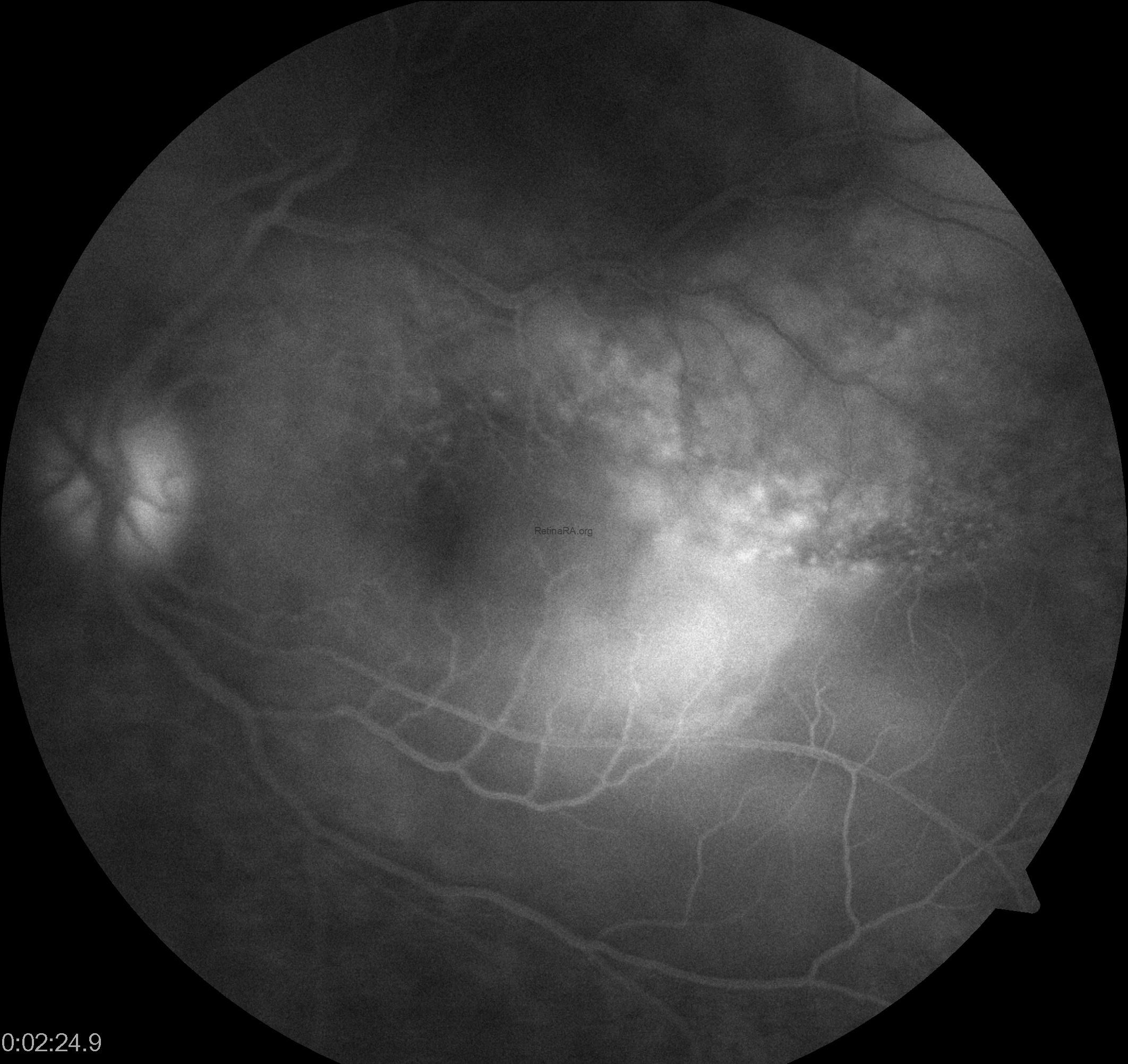
Fluorescein angiography demonstrated optic disc hyperfluorescence, along with pooling and late leakage at the posterior pole.
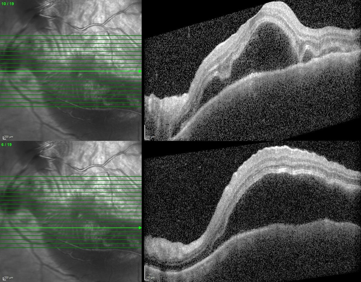
OCT showed marked choroidal thickening, extensive subretinal fluid, and serous retinal detachment.
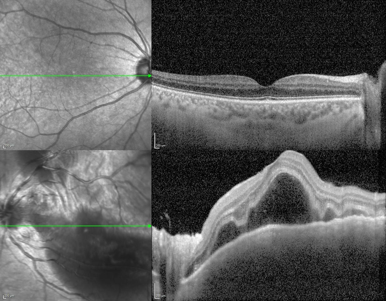
EDI-OCT further demonstrated a markedly thickened choroid in the left eye, with the choroid–sclera interface not clearly discernible.
B-scan ultrasonography revealed posterior scleral thickening with a typical T-sign.
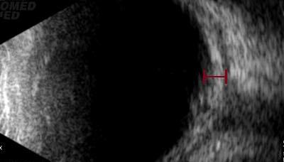
Posterior scleritis is an important masquerader of retinal and choroidal disease. Ocular pain, optic disc edema, disc hyperfluorescence on fluorescein angiography, choroidal folds, exudative retinal detachment, choroidal thickening, and posterior scleral thickening with a T-sign on ultrasonography are important diagnostic clues, even in the absence of anterior chamber or vitreous inflammation.
Credit: Kemal Tekin, M.D., from Ulucanlar Eye Training and Research Hospital
Instagram accounts: @retina.academy and @dr.kemaltekin
