
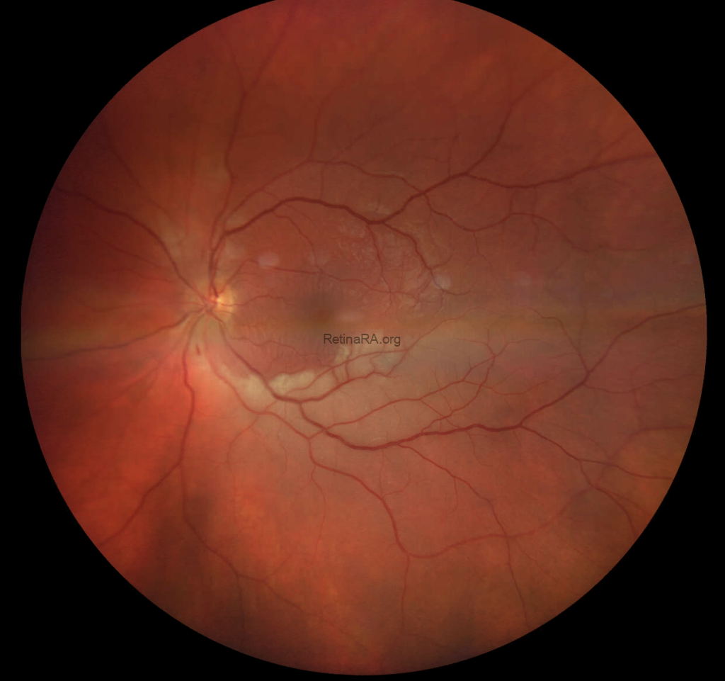
A 36-year-old man presented with an acute superior visual field defect in his left eye that had developed the previous day. Further questioning revealed mild headaches and a subjective reduction in hearing. His medical and surgical histories were unremarkable, and he was not taking any regular medication. Best-corrected visual acuity was 1.25 in both eyes.
Color fundus photography of the right eye was unremarkable. In the left eye, areas of retinal whitening were visible superior to the optic disc and along the inferotemporal vascular arcade, accompanied by flame-shaped hemorrhages. These findings were consistent with branch retinal artery occlusion (BRAO).
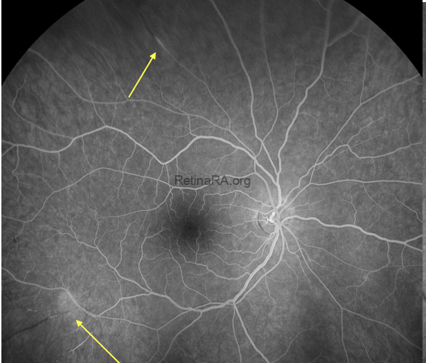
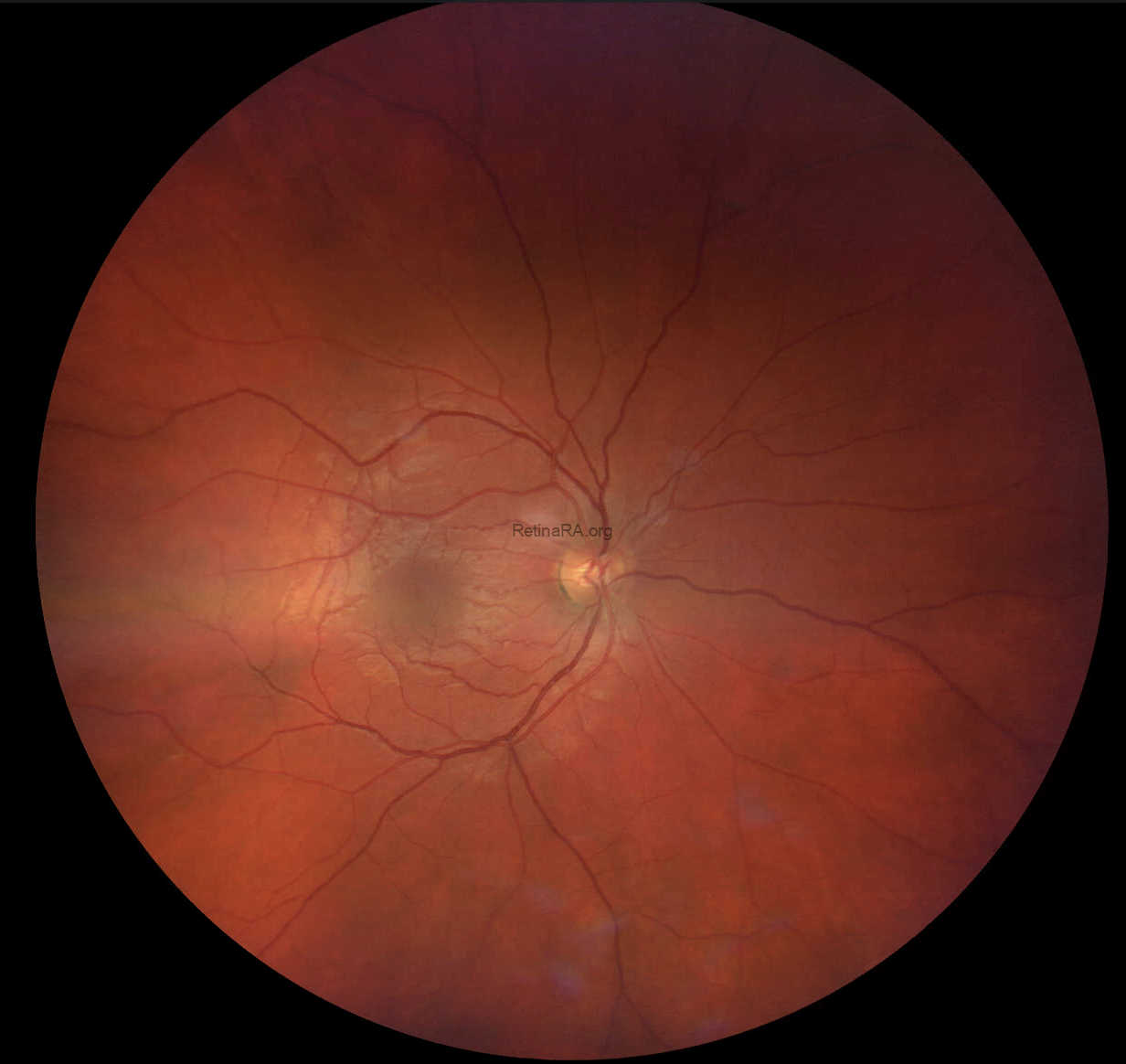 Despite the unremarkable fundus appearance in the right eye, fluorescein angiography (FA) revealed focal arteriolar wall hyperfluorescence with an associated filling defect in the superior retina. These findings demonstrated retinal vascular involvement in the clinically asymptomatic eye.
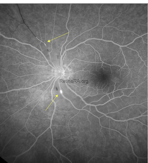
In the symptomatic left eye, FA showed segmental arteriolar wall hyperfluorescence superior to the optic disc, accompanied by a corresponding filling defect. An additional focus of arteriolar wall hyperfluorescence was present inferior to the optic disc.
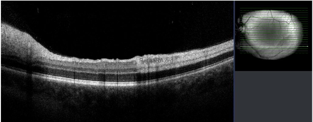
An optical coherence tomography (OCT) scan through the area of retinal whitening along the inferotemporal arcade demonstrated hyperreflectivity of the inner retinal layers, consistent with acute retinal arterial ischemia.
 Brain magnetic resonance imaging (MRI) revealed multiple small lesions involving the corpus callosum, brainstem, including the midbrain, and cerebellum. Audiometry confirmed sensorineural hearing loss. The combination of retinal arterial involvement, cerebral MRI abnormalities, and sensorineural hearing loss supported the diagnosis of Susac syndrome.
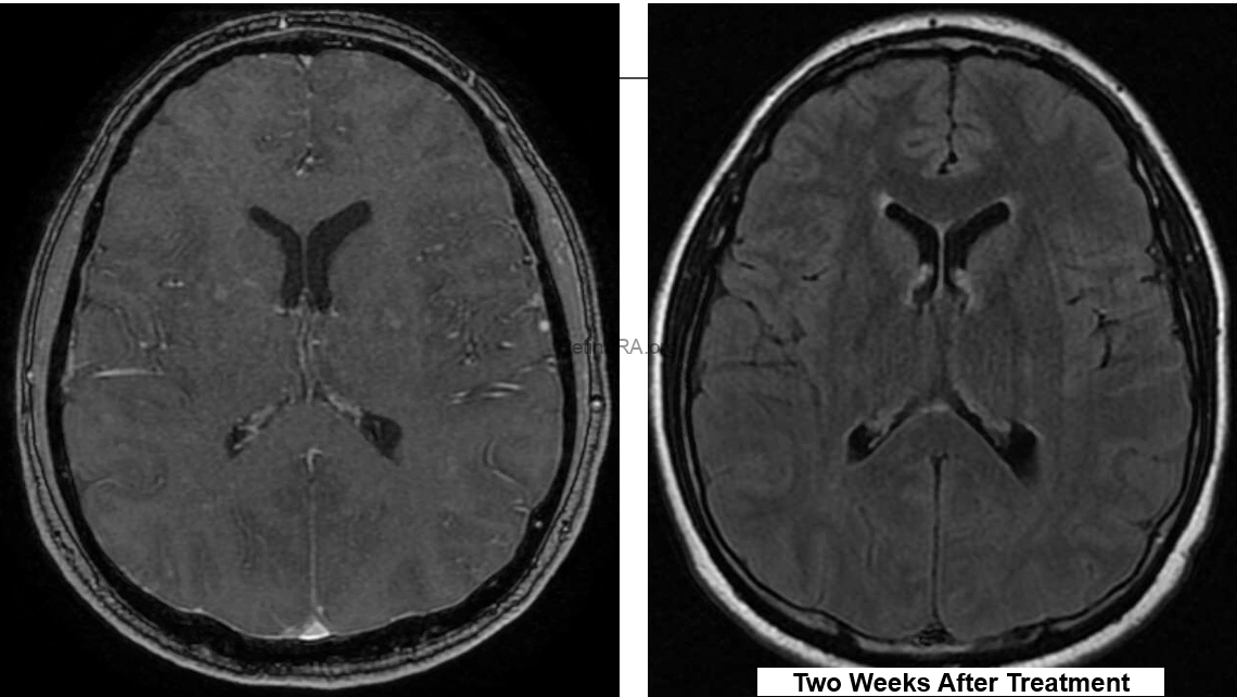 Treatment consisted of high-dose systemic corticosteroids and plasmapheresis. Follow-up brain MRI demonstrated improvement after two weeks of treatment.
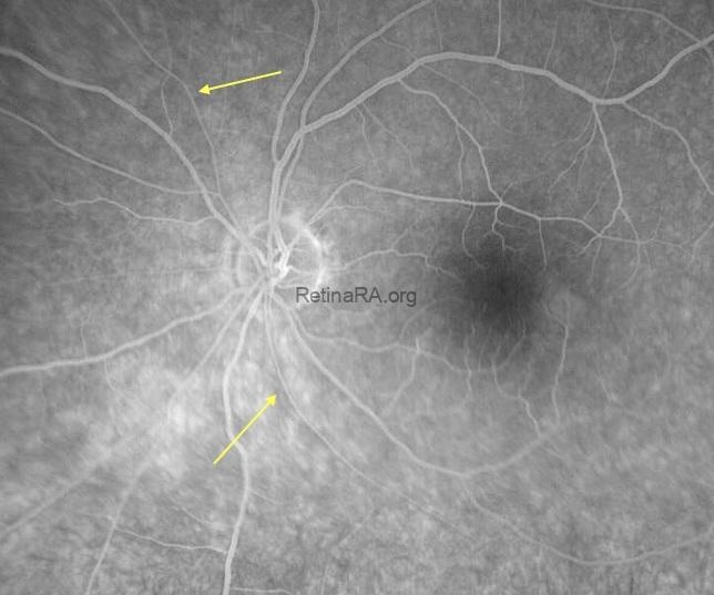
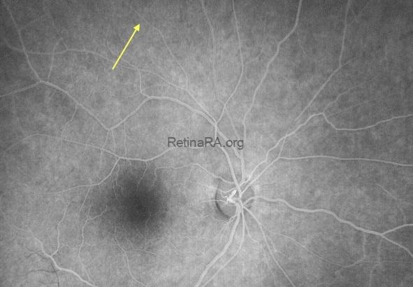
At three months, repeat FA of the left eye showed resolution of arteriolar wall hyperfluorescence at both previously affected sites, with complete reperfusion and no residual angiographic nonperfusion in these areas. Following treatment, fluorescein angiography of the right eye also showed improvement in the previously observed arteriolar abnormalities.
Susac syndrome is a rare immune-mediated microvascular endotheliopathy affecting the brain, retina, and inner ear. It is classically associated with the triad of encephalopathy, branch retinal artery occlusions, and sensorineural hearing loss, although the complete triad may not be apparent at presentation. Retinal findings may provide an early diagnostic clue, particularly in young patients presenting with retinal arterial occlusion accompanied by headaches or auditory symptoms.
This case highlights the value of fluorescein angiography in detecting retinal vascular involvement even when fundus photography appears normal. Recognition of the associated neurological and auditory findings facilitates timely diagnosis and coordinated multidisciplinary care.
Credit: Houssein El Hajjami
Department of Ophthalmology, Klinikum Kassel, Kassel, Germany
Instagram account: @dr.elhajjami
