
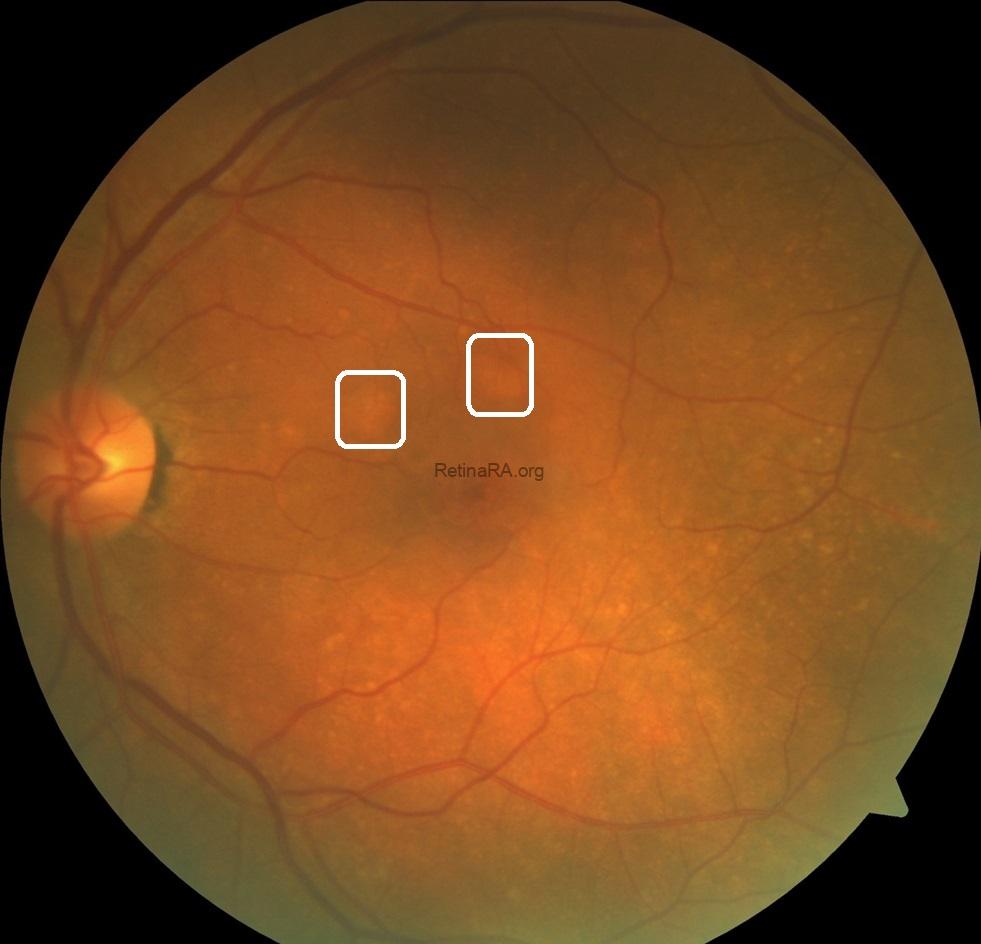
A monocular patient presented for a routine ophthalmic examination. Best-corrected visual acuity was 0.5 in the left eye, and anterior segment examination revealed nuclear cataract.
Fundus examination demonstrated multiple drusen-like deposits and distinct yellow-orange subretinal nodules in the macular region.
 Spectral-domain optical coherence tomography (SD-OCT) revealed characteristic features of polypoidal choroidal vasculopathy (PCV), including sharply peaked pigment epithelial detachments (PEDs), complex multilobular PEDs, and a double-layer sign representing separation of the retinal pigment epithelium from Bruch’s membrane despite the absence of subretinal and intraretinal fluid. The underlying choroid exhibited a pachychoroid configuration with dilated Haller’s layer vessels (pachyvessels).
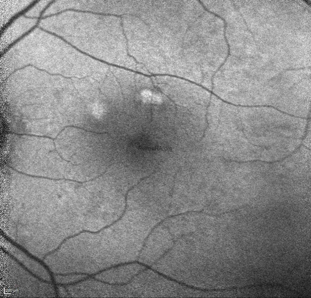
Fundus autofluorescence (FAF) demonstrated focal hyperautofluorescent abnormalities corresponding to the nodular lesions.
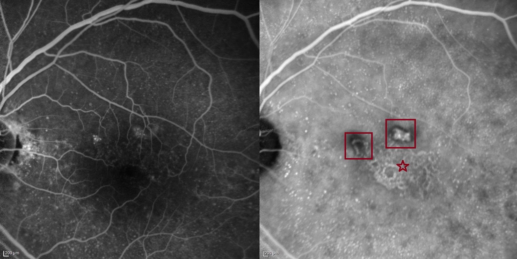 These lesions appeared hyperfluorescent on fluorescein angiography (FA). Indocyanine green angiography (ICGA) revealed hyperfluorescent polypoidal lesions associated with a branching neovascular network, confirming the diagnosis of PCV.
 Optical coherence tomography angiography (OCTA) further delineated the branching neovascular network (BVN) beneath the RPE, demonstrating the vascular architecture of the PCV complex.
This case illustrates the complementary value of multimodal imaging in PCV. While orange subretinal nodules and characteristic OCT findings raise clinical suspicion, ICGA identifies the polypoidal lesions and associated neovascular network, whereas OCTA provides noninvasive visualization of the underlying vascular architecture.
PCV can be clinically categorized into two groups: quiescent (inactive) and active. Quiescent PCV is characterized by the presence of one or more polyps without any intraretinal or subretinal fluid or hemorrhage; it is typically asymptomatic and generally does not require treatment. On the other hand, exudative—or active—PCV manifests through exudation resulting from leakage from active polyps and/or a BVN. This group can be further subdivided into exudative, hemorrhagic, or mixed forms. Clinically, exudation may present as a serous PED, subretinal fluid, serous macular detachment, subretinal lipid exudation, or intraretinal fluid, whereas hemorrhagic PCV manifests as hemorrhage occurring in the subretinal space or beneath the RPE.
Credit: Kemal Tekin, M.D., from Ulucanlar Eye Training and Research Hospital
Instagram accounts: @retina.academy and @dr.kemaltekin
