
Bartonella Neuroretinitis – Case Presentation
A 27-year-old man was referred to our clinic with severe visual loss in his left eye, with visual acuity reduced to counting fingers at 2 meters. Before referral, he had been followed at another center for approximately nine days and had undergone extensive ophthalmological and neurological investigations, including lumbar puncture, but no definitive diagnosis had been established and no treatment had been initiated.
Initial Presentation to Our Clinic
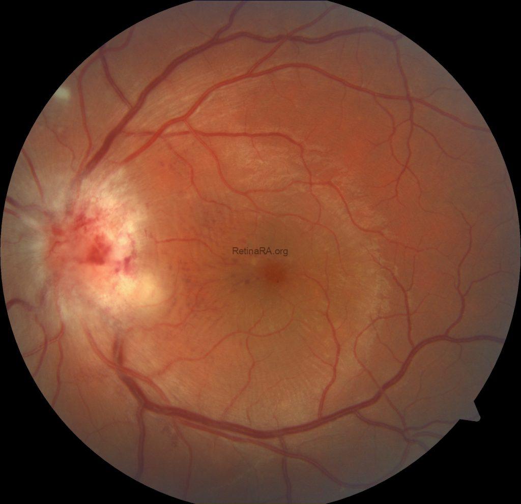
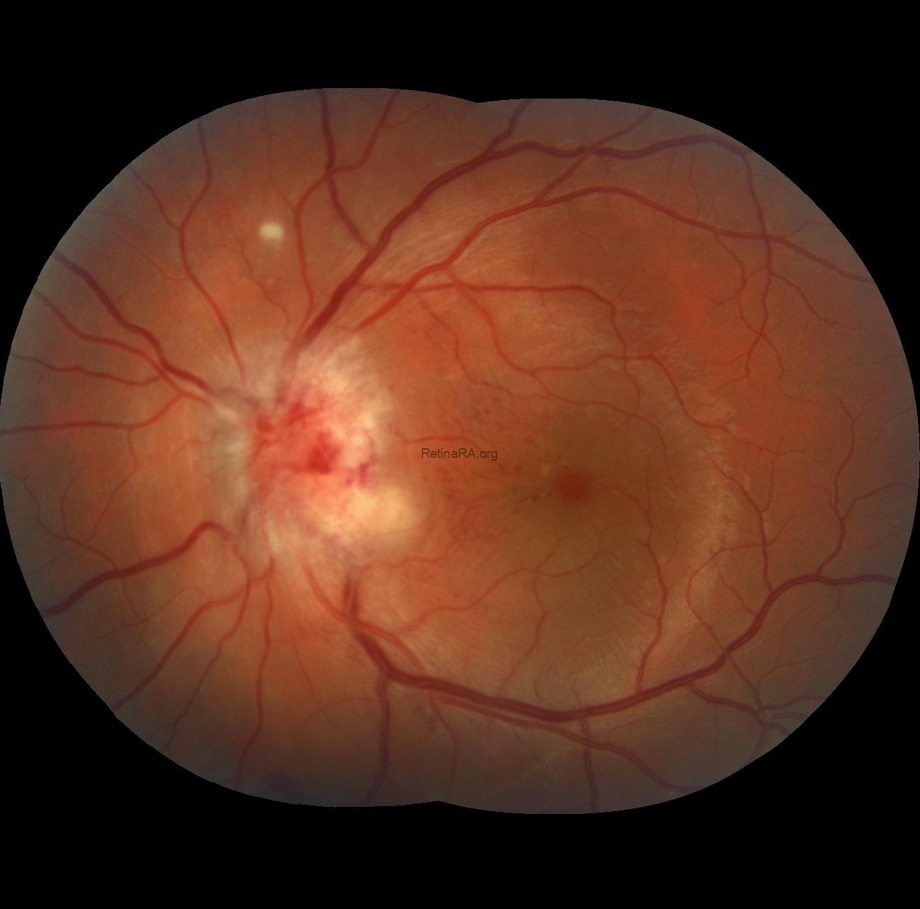
At his first examination in our clinic, color fundus photography revealed marked optic disc edema associated with peripapillary hemorrhages. Extensive subretinal fluid extended from the optic disc toward the macula and surrounding retina.
In addition, a separate retinitis focus was visible approximately two disc diameters superior to the optic disc. Another whitish area of retinitis could be identified inferotemporal to the swollen optic disc, extending toward the papillomacular region. Because of the severe optic disc edema and extensive retinal involvement, the borders of these inflammatory lesions were not sharply demarcated.
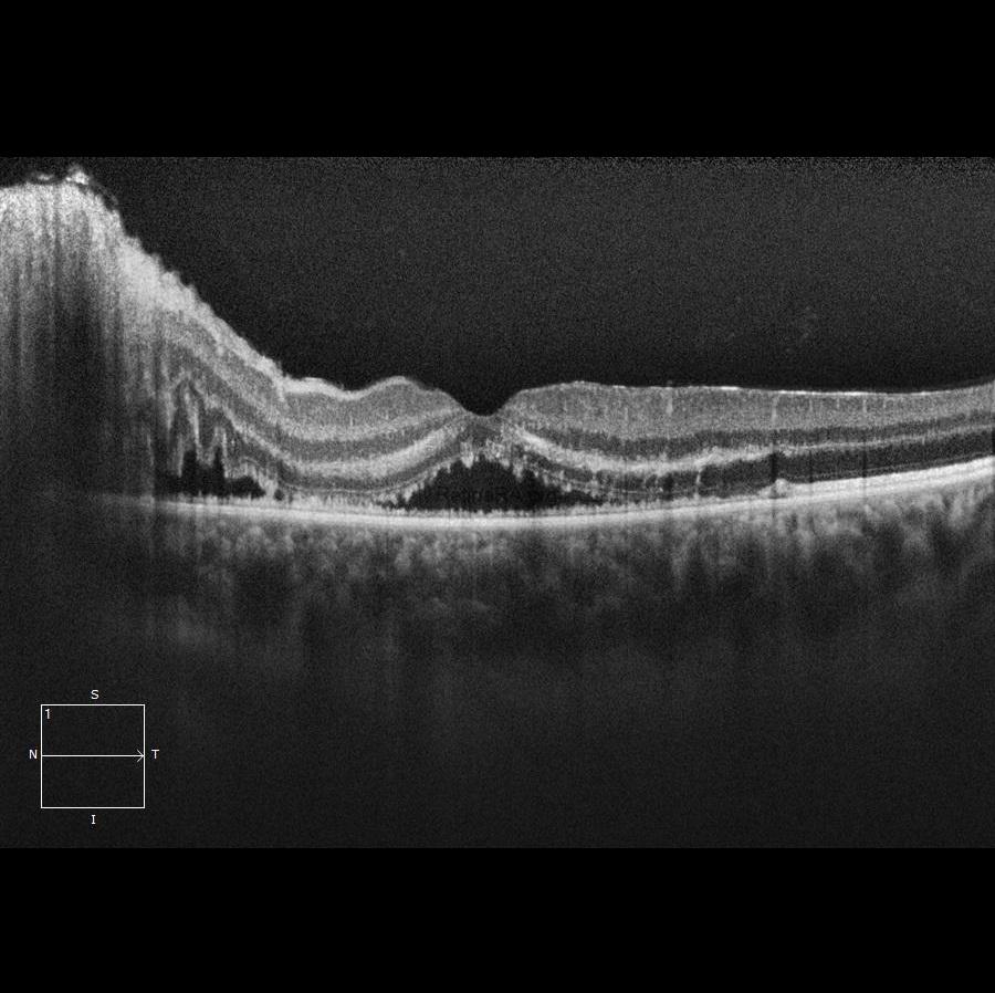
OCT confirmed the extensive subretinal fluid involving the macula and marked swelling of the optic nerve head.

Despite the absence of a history of cat ownership or known cat contact, the clinical findings strongly suggested Bartonella neuroretinitis. Therefore, without waiting for serologic confirmation, treatment with oral doxycycline, rifampicin, and systemic prednisolone was initiated immediately.
Day 4
A rapid clinical response was observed. By the fourth day of treatment, optic disc edema and hemorrhages had already decreased, and OCT demonstrated a clear reduction in subretinal fluid.
Interestingly, the characteristic macular star became more apparent as the subretinal fluid resolved. Although the patient had already been symptomatic for approximately 9–10 days before presentation to our clinic, the macular star was initially poorly visible because of the extensive fluid accumulation. As the fluid decreased, the typical stellate pattern of macular deposits gradually emerged.

Day 13
By Day 13, further regression of the optic disc edema and retinitis lesions was evident. Yellowish deposits became increasingly prominent in the areas previously occupied by subretinal fluid, producing a more characteristic macular star appearance.
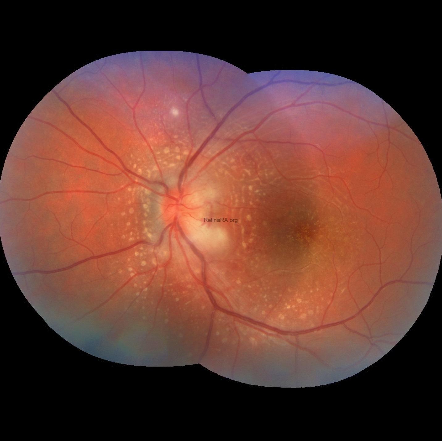

The retinitis foci progressively decreased in size, accompanied by continued anatomical improvement on OCT.
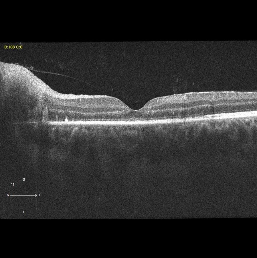
Serologic testing subsequently demonstrated positive Bartonella henselae IgM, supporting the clinical diagnosis. Investigations for alternative infectious causes, including Borrelia and toxoplasmosis, were negative.
One-Month Follow-up:
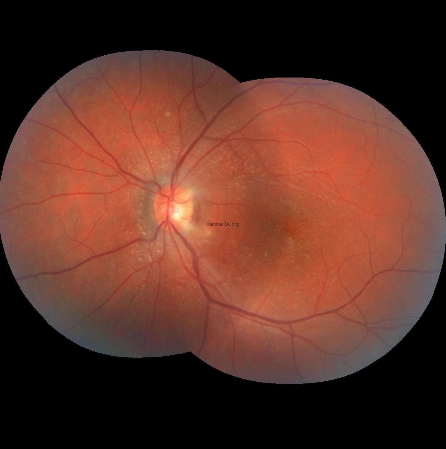
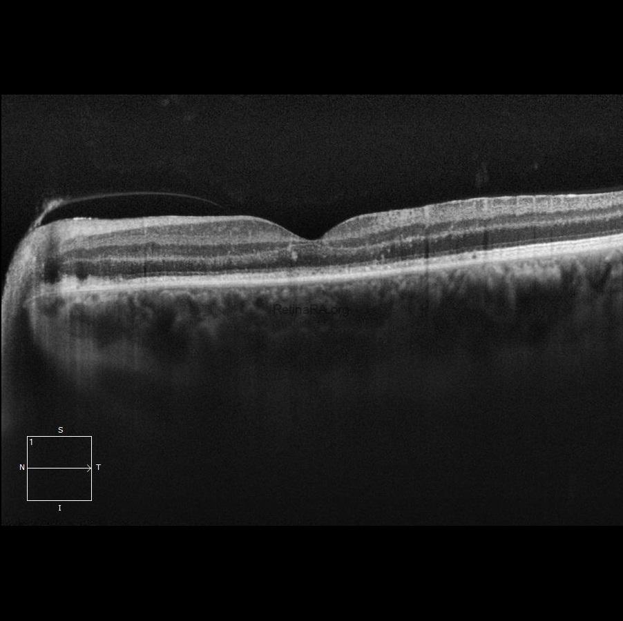
At the one-month visit, the optic disc edema had essentially resolved and the yellowish retinal deposits had markedly decreased. Mild peripapillary atrophic changes, more prominent on the nasal side of the optic disc, had begun to develop. OCT showed complete resolution of the subretinal fluid; however, disruption of the outer retina and ellipsoid zone, particularly at the fovea, remained evident. Visual acuity had improved to 0.1.
Day 45 Follow-up:
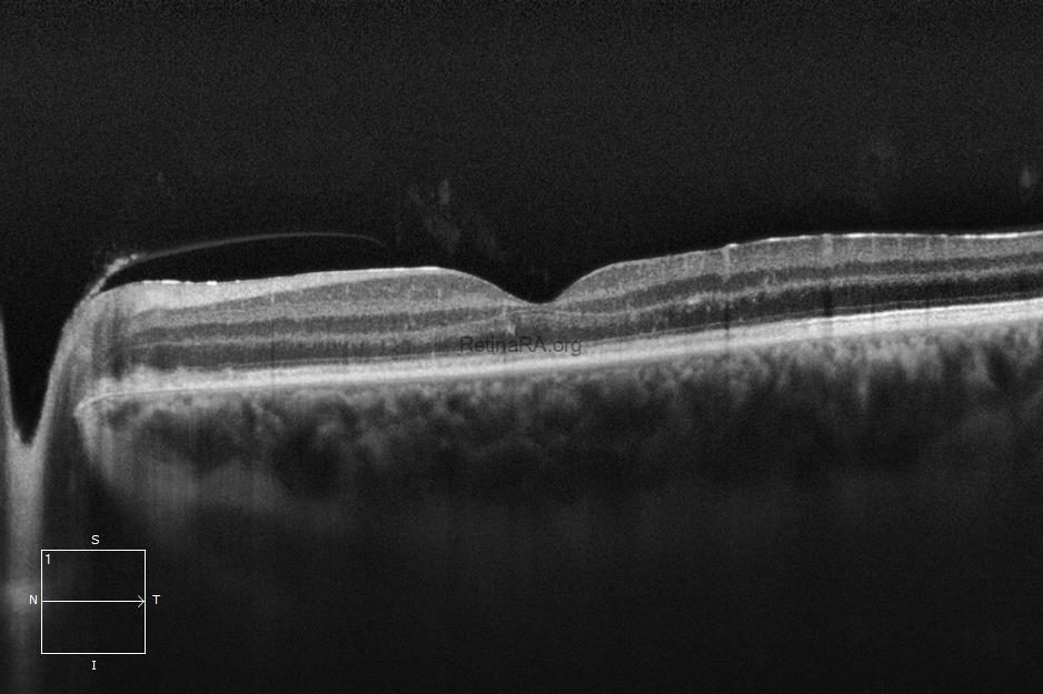
By Day 45, the retinal deposits had further regressed, with no recurrence of optic disc edema or subretinal fluid. OCT demonstrated progressive restoration of the photoreceptor layers and ellipsoid zone. This anatomical recovery was accompanied by further functional improvement, with visual acuity increasing to 0.3.

Bartonella neuroretinitis classically presents with optic disc edema accompanied by subretinal fluid, followed by the development of a macular star. However, the macular star may not be apparent at initial presentation and typically becomes more evident several days later. In severe cases with extensive subretinal fluid, as in our patient, the characteristic stellate pattern may initially be obscured and become visible only as the fluid resolves. Importantly, a history of cat exposure is not always identified, and negative serologic testing does not completely exclude the diagnosis, particularly early in the disease course, when Bartonella-specific IgM or other serologic tests may still be negative. Therefore, the diagnosis should be based on the overall clinical picture rather than on a single historical or laboratory finding.
For mild cases of Bartonella neuroretinitis in which antimicrobial treatment is considered appropriate, doxycycline may be used as monotherapy, whereas in severe or vision-threatening cases, particularly those with marked optic disc or macular involvement, doxycycline combined with rifampin is commonly preferred. Systemic corticosteroids may be considered as adjunctive therapy in patients with severe optic disc inflammation, extensive subretinal fluid, or significant macular involvement and visual loss. In such severe cases, corticosteroids may be initiated concomitantly with appropriate antibiotic therapy, although some clinicians prefer to introduce systemic corticosteroids several days after starting antimicrobial treatment. The optimal treatment regimen and timing of corticosteroid therapy remain controversial, as current evidence is largely based on retrospective studies and case series.
Credit: M. Giray Ersoz, MD, FEBO, Retina Specialist
Memorial Bahçelievler Hospital, Department of Ophthalmology, Istanbul, Turkey
Arel University School of Medicine, Department of Ophthalmology, Istanbul, Turkey
Instagram accounts: @retina.review and @retina.dr.girayersoz
Website: www.girayersoz.com.tr
