

A 35-year-old man presented with central serous chorioretinopathy (CSC) in his left eye. His right eye was completely asymptomatic, with a best-corrected visual acuity of 20/20. However, multimodal imaging of the asymptomatic right eye revealed findings consistent with pachychoroid pigment epitheliopathy (PPE).
On optical coherence tomography (OCT), several focal abnormalities were observed at the level of the retinal pigment epithelium (RPE), indicated by white arrows. PPE lesions may exhibit different morphologic patterns on OCT, including focal RPE thickening, small pigment epithelial detachments, microbreak-like RPE elevations or hyperreflective RPE spikes, and hyperreflective RPE columns. In this case, the lesions predominantly appeared as focal RPE thickening and small RPE detachments. OCT also demonstrated prominent pachyvessels within the choroid, further supporting the underlying pachychoroid phenotype.


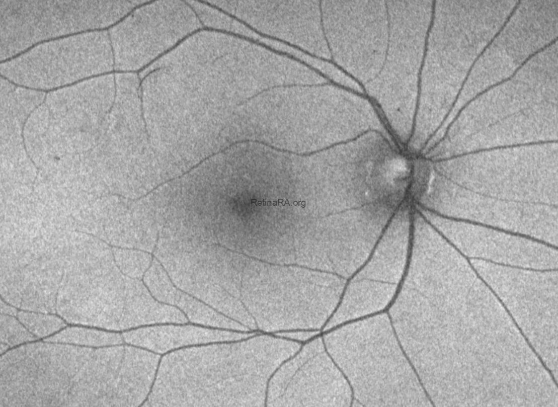
Fundus autofluorescence (FAF) imaging did not demonstrate significant abnormalities. In particular, there were no characteristic autofluorescence changes suggesting previous episodes of CSC or extensive RPE damage. Together with the absence of symptoms and previous subretinal fluid, these findings supported the diagnosis of PPE rather than previously resolved CSC.
Fluorescein angiography (FA), which was performed because of active CSC in the fellow eye, allowed simultaneous evaluation of the asymptomatic right eye. FA demonstrated several small hyperfluorescent lesions located predominantly along the temporal portions of the superior and inferior vascular arcades. These lesions, indicated by red arrows, corresponded spatially to the RPE abnormalities identified on OCT with white arrows.

Indocyanine green angiography (ICGA) provided further information regarding the underlying choroidal circulation. In the early phase, asymmetric venous drainage was observed, with prominent drainage through the inferotemporal vortex vein. During the mid-to-late phases of ICGA, multiple areas of choroidal vascular hyperpermeability became evident as hypercyanescent areas, indicated by green arrows. Hyperpermeability was present not only beneath the areas corresponding to the visible PPE lesions but also in additional regions without clearly detectable RPE lesions

Ultra-widefield OCT angiography was additionally used to evaluate the distribution of choroidal vascular volume (CVV). The CVV map demonstrated a prominent increase in choroidal vascular volume within the central choroid, with an asymmetric distribution extending from the inferotemporal vortex vein territory toward the inferotemporal macular region and superiorly.

What Is Pachychoroid Pigment Epitheliopathy?
Pachychoroid pigment epitheliopathy represents an early and frequently asymptomatic manifestation of the pachychoroid disease spectrum. It is characterized by focal RPE abnormalities occurring in eyes with an underlying pachychoroid phenotype, without current or previous subretinal fluid. The condition is therefore particularly important in the asymptomatic fellow eyes of patients presenting with unilateral CSC. In our previous study of 282 patients with unilateral CSC, PPE was identified in 61% of asymptomatic fellow eyes, whereas 30.8% had uncomplicated pachychoroid and only 8.2% had normal fellow eyes.[2]
The abnormalities visible at the RPE level represent only one component of a more extensive choroidal disorder. ICGA studies have demonstrated that PPE is associated with choroidal vascular hyperpermeability, supporting the concept that the primary pathophysiologic alterations occur within the choroidal circulation.[3] PPE lesions themselves may also evolve over time. They may appear as focal RPE thickening, pigment epithelial detachment, or microbreak-like RPE elevations, and some lesions can subsequently progress to CSC. In our long-term study, 17.4% of eyes with PPE developed CSC during follow-up, supporting the concept that PPE may represent a precursor phenotype within the pachychoroid disease spectrum.[4]
The present case illustrates this concept particularly well: although the right eye was asymptomatic, had 20/20 vision, and showed no evidence of previous CSC on FAF, multimodal imaging revealed prominent pachyvessels, focal PPE lesions, asymmetric vortex vein drainage, widespread choroidal vascular hyperpermeability, and increased choroidal vascular volume. Thus, the visible RPE lesions may represent the localized retinal manifestation of a substantially broader underlying choroidal vascular abnormality.
Credit: M. Giray Ersoz, MD, FEBO, Retina Specialist
Memorial Bahçelievler Hospital, Department of Ophthalmology, Istanbul, Turkey
Arel University School of Medicine, Department of Ophthalmology, Istanbul, Turkey
Instagram accounts: @retina.review and @retina.dr.girayersoz
Website: www.girayersoz.com.tr
References
- Warrow DJ, Hoang QV, Freund KB. Pachychoroid pigment epitheliopathy. Retina. 2013;33(8):1659–1672. doi:10.1097/IAE.0b013e3182953df4.
- Ersoz MG, Karacorlu M, Arf S, Hocaoglu M, Sayman Muslubas I. Pachychoroid pigment epitheliopathy in fellow eyes of patients with unilateral central serous chorioretinopathy. Br J Ophthalmol. 2018;102(4):473–478. doi:10.1136/bjophthalmol-2017-310724.
- Ersoz MG, Arf S, Hocaoglu M, Sayman Muslubas I, Karacorlu M. Indocyanine green angiography of pachychoroid pigment epitheliopathy. Retina. 2018;38(9):1668–1674. doi:10.1097/IAE.0000000000001773.
- Karacorlu M, Ersoz MG, Arf S, Hocaoglu M, Sayman Muslubas I. Long-term follow-up of pachychoroid pigment epitheliopathy and lesion characteristics. Graefes Arch Clin Exp Ophthalmol. 2018;256(12):2319–2326. doi:10.1007/s00417-018-4144-0.
- Ersoz MG, Karacorlu M, Arf S, Hocaoglu M, Sayman Muslubas I. Outer nuclear layer thinning in pachychoroid pigment epitheliopathy. Retina. 2018;38(5):957–961. doi:10.1097/IAE.0000000000001655.
