
A 15-year-old girl was referred for reduced vision and strabismus in her right eye. Best-corrected visual acuity was 0.4 in the right eye and 1.0 in the left eye. Anterior segment examination was unremarkable in both eyes.
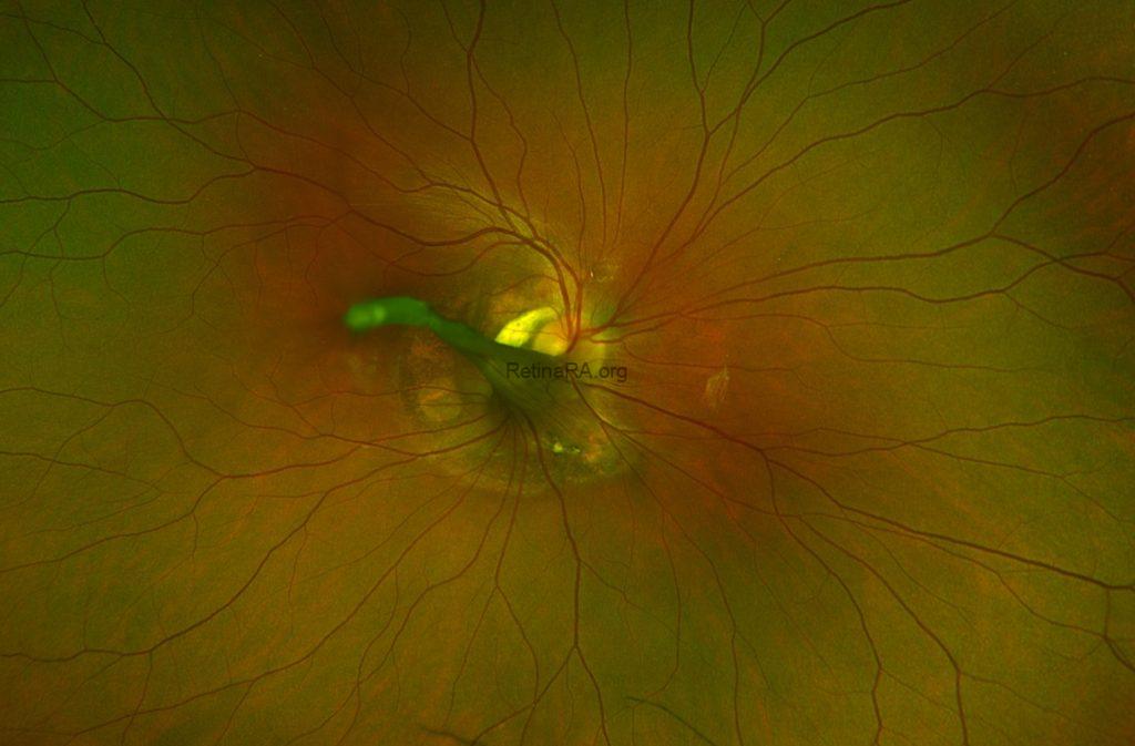
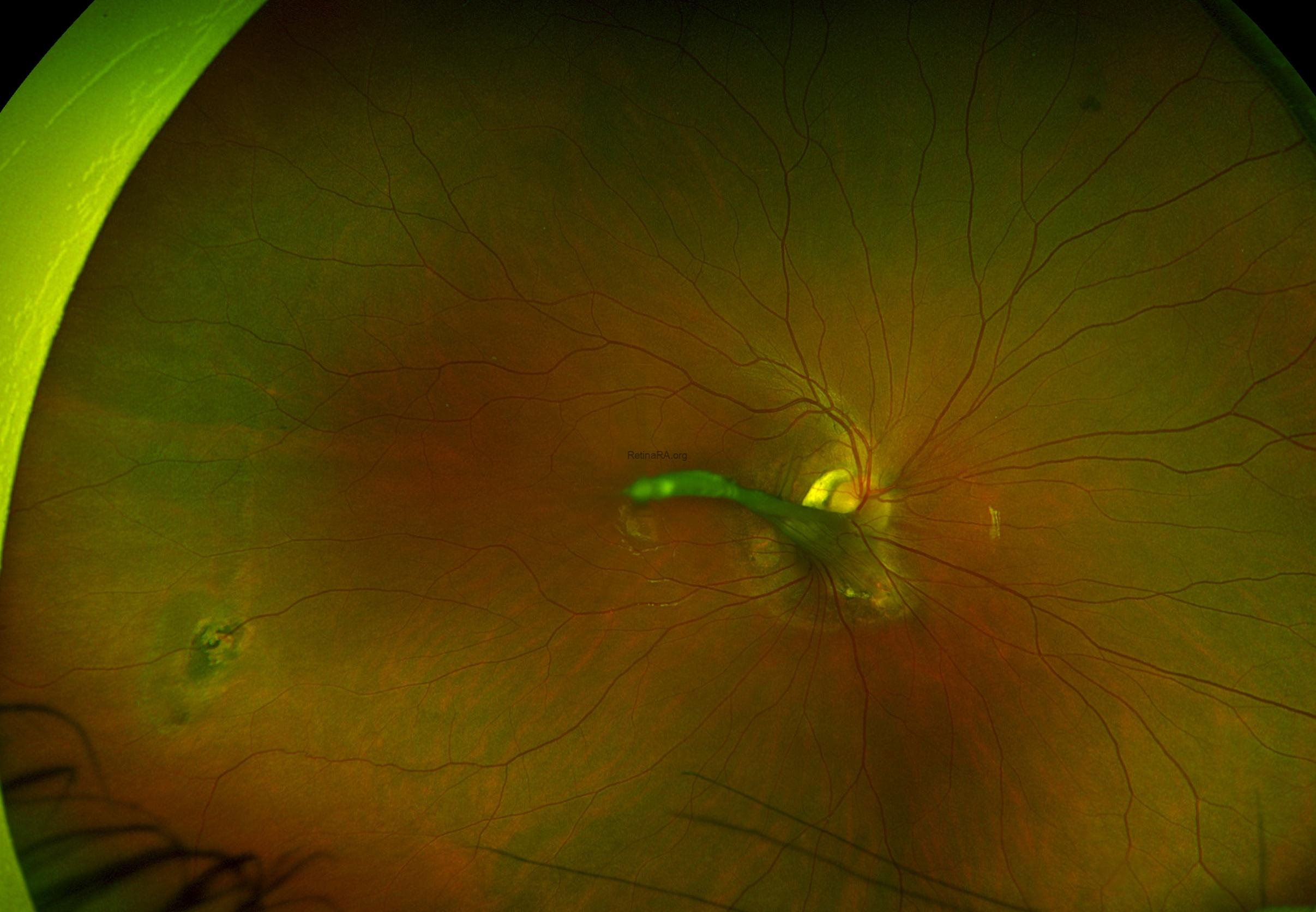
Ultra-widefield fundus imaging of the right eye revealed a prominent fibrovascular stalk extending anteriorly from the optic nerve head into the vitreous cavity, associated with vitreoretinal traction and retinal dragging toward the optic disc. Peripapillary retinal folds and localized chorioretinal pigmentary alterations were also observed. A separate peripheral chorioretinal lesion was visible in the inferotemporal retina.
The fellow eye had normal posterior segment structures.

Fundus autofluorescence of the right eye demonstrated irregular areas of hypo- and hyperautofluorescence surrounding the optic disc.

These findings were consistent with posterior persistent fetal vasculature (PFV).
Persistent fetal vasculature, previously known as persistent hyperplastic primary vitreous, is a congenital developmental anomaly resulting from incomplete regression of the fetal hyaloid vascular system. Most cases are sporadic, without an identifiable genetic cause. PFV is an important cause of childhood leukocoria and amblyopia. While anterior PFV typically presents with retrolental fibrovascular tissue and lens abnormalities, posterior PFV is characterized by a fibrovascular stalk arising from the optic disc, retinal dragging, tractional folds, and variable macular involvement.
Credit: Kemal Tekin, M.D., from Ulucanlar Eye Training and Research Hospital
Instagram accounts: @retina.academy and @dr.kemaltekin
