
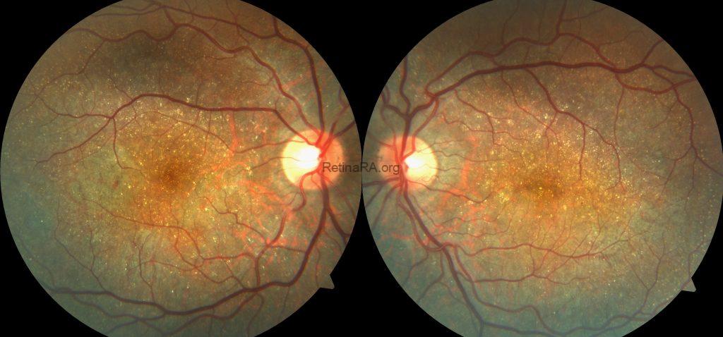
A 30-year old male patient applied with routine ocular examination. Best-corrected visual acuity was 0.8 in both eyes, and anterior segment examination was unremarkable bilaterally.
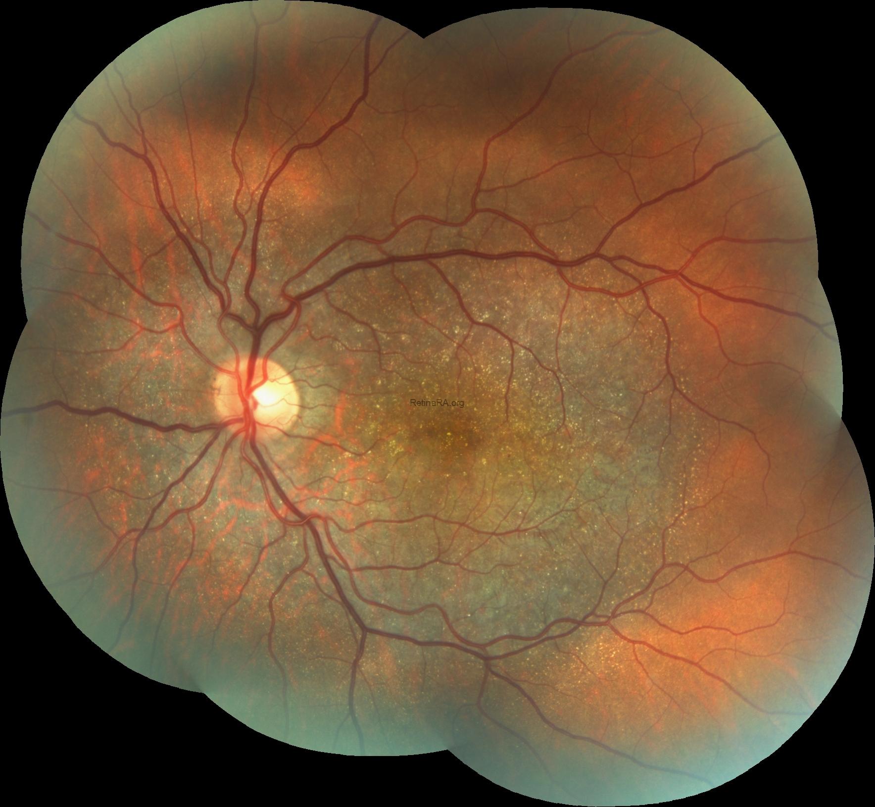
Color fundus images revealed numerous tiny, glistening yellow-white crystalline deposits scattered throughout the posterior pole and extending into the midperipheral retina in both eyes. Diffuse retinal pigment epithelium alterations and chorioretinal atrophic changes were also evident, with increased visibility of the underlying choroidal vessels.
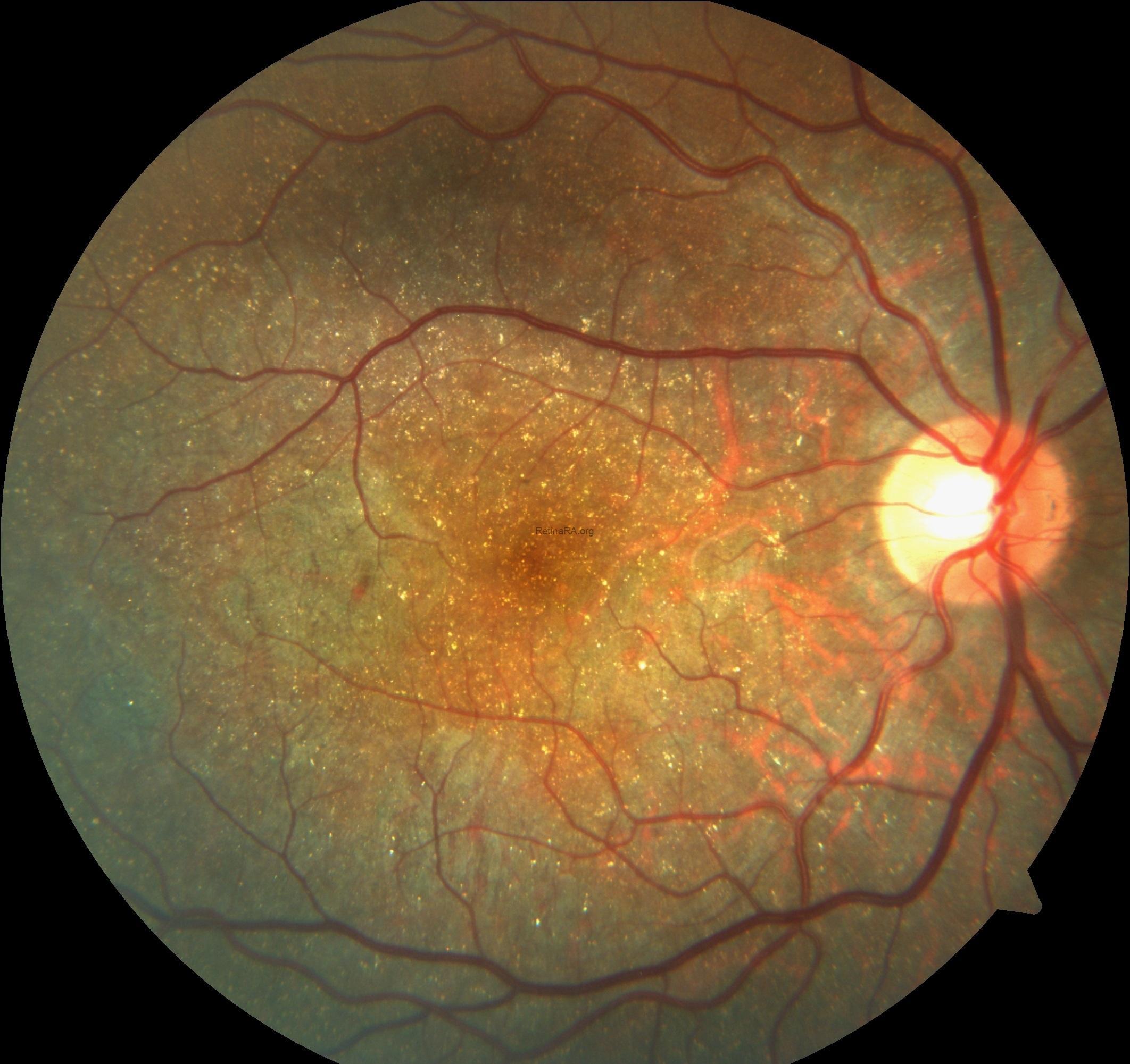
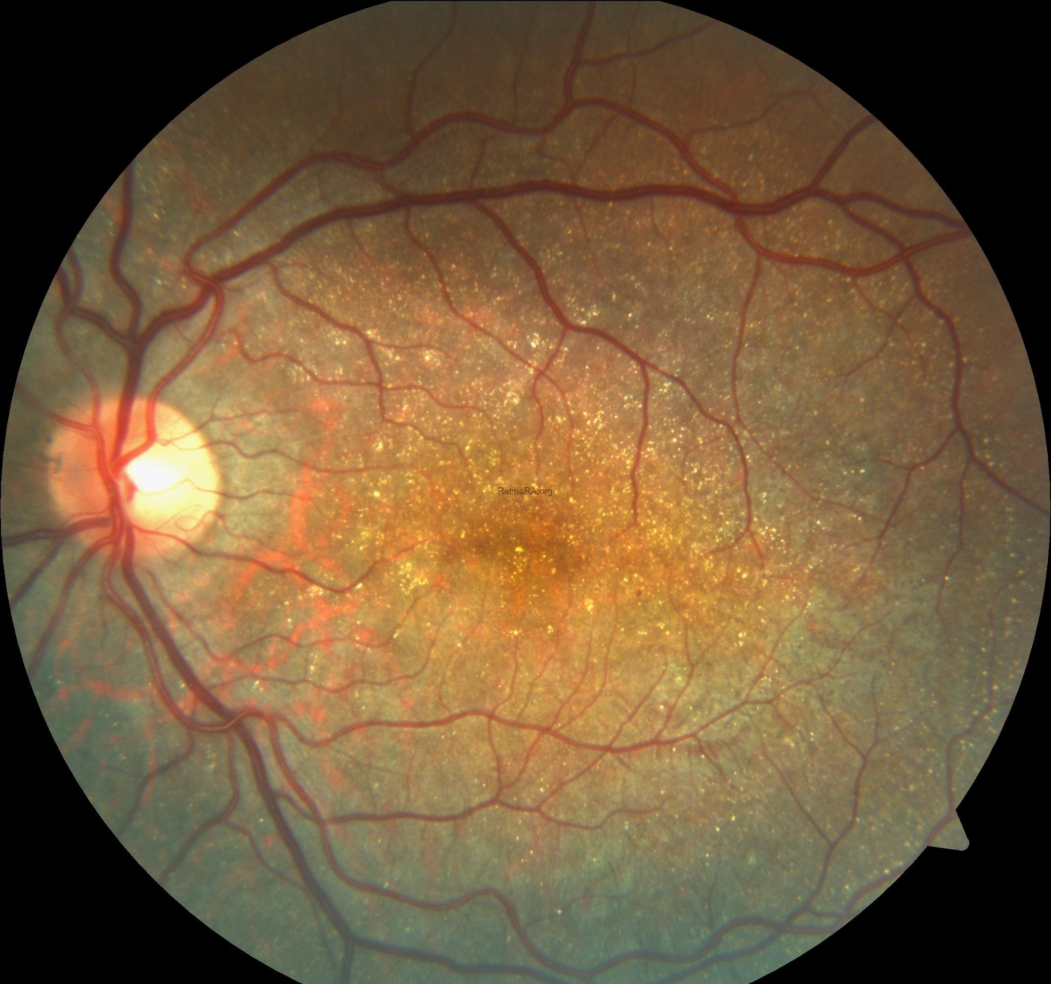
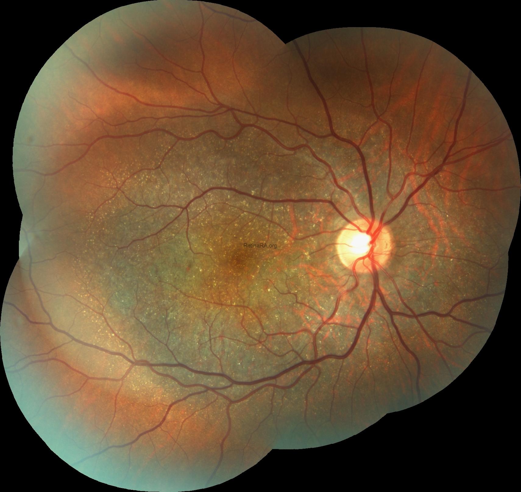
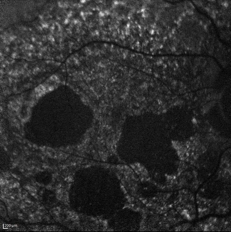
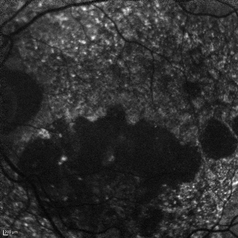
Fundus autofluorescence demonstrated bilateral, irregular, well-demarcated areas of markedly reduced autofluorescence, corresponding to RPE atrophy. These hypoautofluorescent lesions were surrounded by a heterogeneous, granular autofluorescence pattern, reflecting widespread RPE alterations.
Spectral-domain optical coherence tomography showed bilateral RPE irregularities, focal outer retinal attenuation, and scattered punctate hyperreflective foci. Despite the extensive chorioretinal changes, the foveal contour and central outer retinal architecture were relatively preserved, consistent with the maintained central visual acuity.
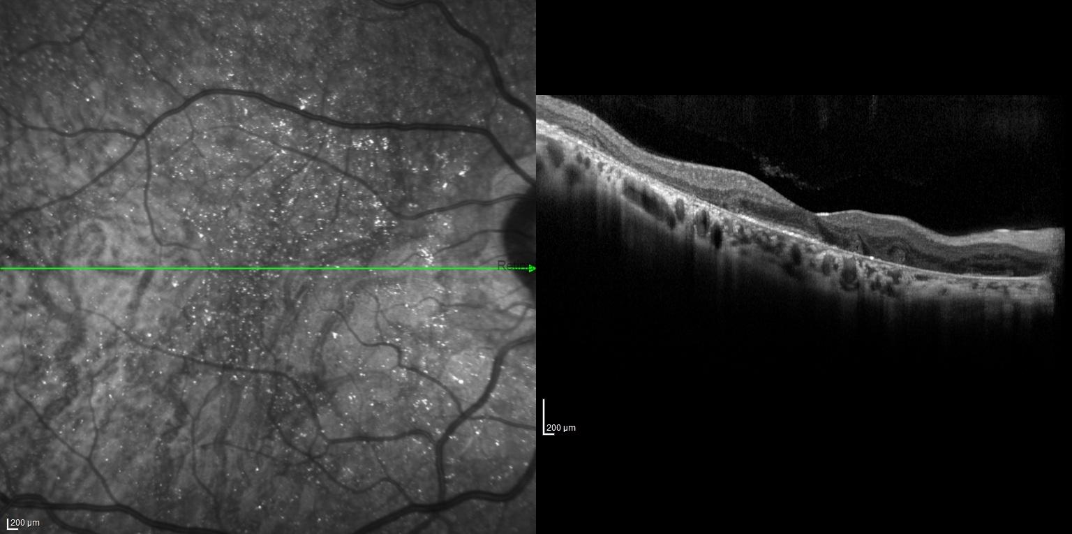
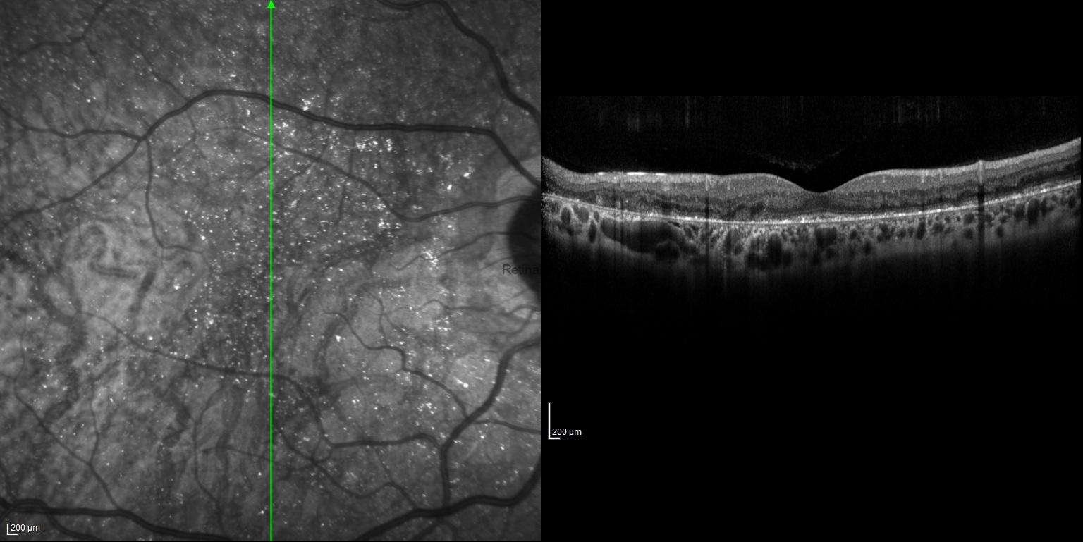
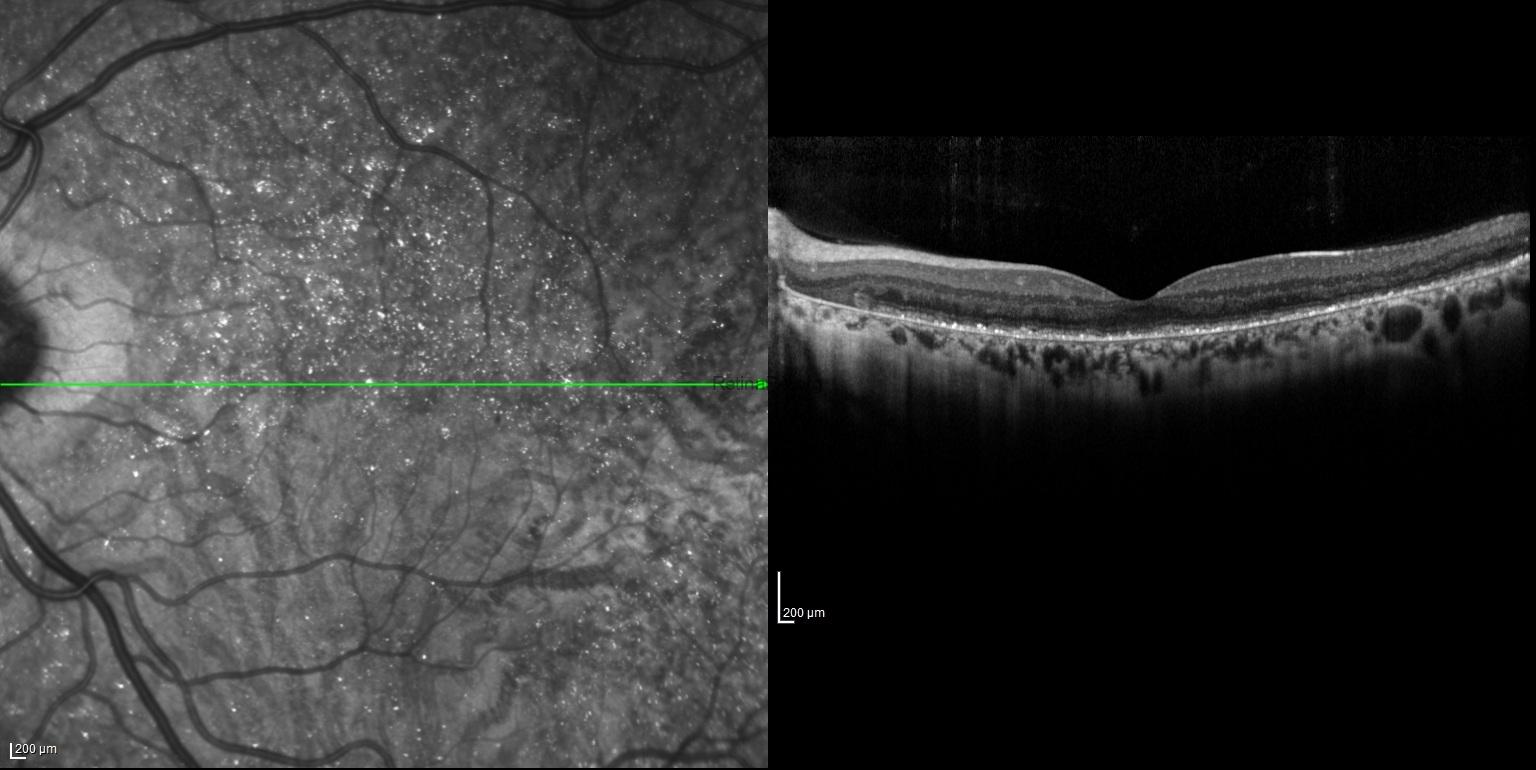
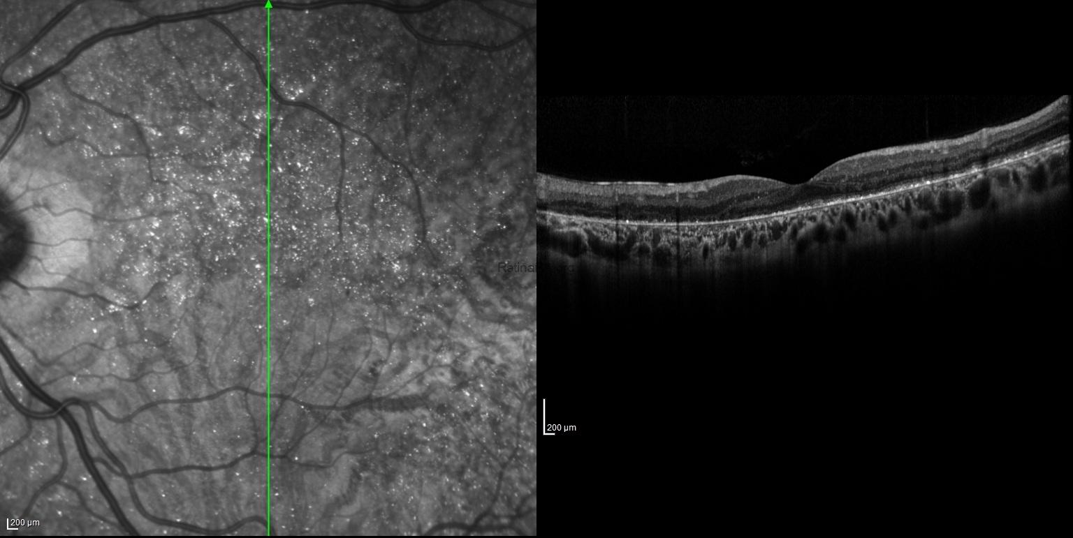
Bietti crystalline dystrophy is a rare, autosomal recessive chorioretinal degeneration associated with biallelic pathogenic variants in the CYP4V2 gene. It is characterized by intraretinal crystalline deposits, progressive RPE and choriocapillaris atrophy, and outer retinal degeneration. Crystalline deposits may become less prominent as the disease progresses. Multimodal imaging is essential for evaluating disease extent and monitoring progression. Relative preservation of the foveal photoreceptor structure may allow good central visual acuity despite extensive surrounding chorioretinal atrophy.
Credit: Kemal Tekin, M.D., FEBOS-R, from Ulucanlar Eye Training and Research Hospital
Instagram accounts: @retina.academy and @dr.kemaltekin
