

The case describes a 4-year-old male patient who presented with a negative general and ocular history. Upon examination, best-corrected visual acuity (VA) was 20/20 in both eyes (OU), and the anterior segment was unremarkable. The patient underwent a comprehensive multimodal imaging evaluation.



Discussion and Pathogenetic Considerations
Stellate Multiform Amelanotic Choroidopathy (SMACH), originally described as Serous Maculopathy due to Aspecific Choroidopathy, is an uncommon choroidal disorder with characteristic multimodal imaging findings. The revised terminology encompasses cases both with and without subretinal fluid.
Clinical and Multimodal Imaging Findings
- Fundus Appearance: SMACH typically presents as a unilateral, amelanotic choroidal lesion with irregular, stellate or branching margins. Published series describe a generally stable course without documented lesion enlargement during follow-up.
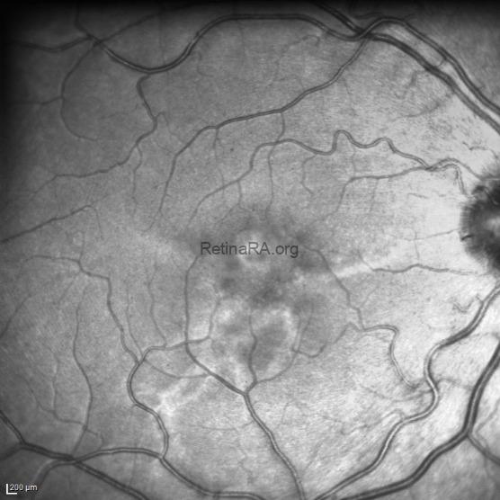
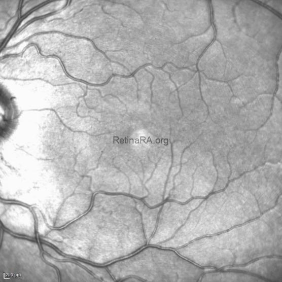
- Findings in the Present Case and Interocular Comparison: Color fundus photography shows a pale yellowish lesion with irregular, branching margins in the macular region. The lesion is particularly conspicuous on infrared reflectance imaging, which highlights its irregular hyperreflective margins and branching extensions. Comparison with the fellow eye demonstrates no corresponding lesion or similar reflectance pattern in the imaged macular region. The fellow-eye infrared image is included as a reference to emphasize this interocular asymmetry and the unilateral appearance of the findings.
- Cross-sectional OCT: Infrared-guided and color fundus–guided OCT scans demonstrate hyperreflective changes within the inner choroid, accompanied by mild contour alterations of the overlying RPE–Bruch’s membrane complex. The foveal contour is preserved, and no definite intraretinal or subretinal fluid is evident in the displayed sections.
- OCT Angiography and En Face OCT: The upper panels display OCT angiography at different segmentation levels, while the lower panels show the corresponding en face structural images. Horizontal motion artifacts limit detailed interpretation of the angiographic findings; however, no evidence of macular neovascularization is identified in the available images.
Key Literature Highlights
- Subretinal Fluid: SRF is a variable feature rather than a requirement for diagnosis. It was present in 10 of 18 eyes (56%) at presentation in the original multicenter series, whereas eight eyes remained free of SRF during follow-up. Fluid may persist, fluctuate or resolve spontaneously.
- Macular Neovascularization: MNV was not identified in the cited SMACH series. This observation should not be interpreted as proof that neovascularization can never occur.
- Treatment Response: Early reports described limited responses to photodynamic therapy and intravitreal anti-VEGF injections. Subsequent case reports have documented SRF resolution after PDT in selected patients (Van Gorcom et al). Evidence remains limited, and a consistently effective treatment strategy has not been established.
- Pathogenesis: The underlying mechanism remains uncertain. Early detection and reported lesion stability suggest a possible developmental abnormality of the choroid. Local effects on the choriocapillaris and overlying RPE may contribute to fluid accumulation, potentially through altered hydrostatic pressure and impaired barrier function. These mechanisms remain hypotheses.
Differential Diagnosis
Given the clinical presentation, the diagnostic workup required ruling out several conditions, including:
-
Choroidal tumors
-
Chronic central serous chorioretinopathy (CSC)
-
Inherited retinal disorders
-
Inflammatory chorioretinopathies
Key Takeaway & Clinical Note:
While previous literature reports the youngest patient detected with this condition at 7 years of age, our 4-year-old patient represents the youngest case described in the literature to date, further strengthening the hypothesis of a congenital choroidal dysgenesis.
Credit:
Giovanni Di Fiore, M.D. Naples @giodifiore
Claudio Iovino, M.D. Naples @claudioiovino
Key Bibliographic References:
-
Ramtohul P, Pellegrini M, Pichi F, Preziosa C, Marchese A, Cicinelli MV, Miserocchi E, Mundae R, Mrejen S, Rofagha S, Mein CE, Mein L, Ober MD, Cunha de Souza E, Cohen SY, van Dijk EHC, Jampol L, Boon CJF, Freund KB.Stellate Multiform Amelanotic Choroidopathy (SMACH). Clinical and Multimodal Imaging Features. Retina. 2023 Sep 1;43(9):1532-1540.
-
van Dijk EHC, Boon CJF, et al.Serous Maculopathy Due to Aspecific Choroidopathy (SMACH). (Described within the differential diagnosis spectrum of central serous chorioretinopathy and pachychoroid disease spectrum).
-
van Dijk EHC, Boon CJF.Serous business: Delineating the broad spectrum of diseases with subretinal fluid in the macula. Progress in Retinal and Eye Research. 2021 Sep;84:100955.
-
Jeon J, Kwak JJ, Seoung HJ, Kim J, Byeon SH, Kim SS, Woo SJ, Lee CS. Clinical and imaging features of SMACH. BMC Ophthalmol. 2026 Apr 14;26(1):281. doi: 10.1186/s12886-026-04810-z. PMID: 41981541; PMCID: PMC13188576.
-
Arango-Hurtado M, Quintero-Lizcano K, Díaz-Vargas JA, Mejía ST, Velásquez SR. SMACH: A Rare, Non-Progressive Retinal Disorder – Key Insights into Its Clinical and Imaging Characteristics. A Case Report. Rom J Ophthalmol. 2026 Apr-Jun;70(2):326-329. doi: 10.22336/rjo.2026.44. PMID: 42572626; PMCID: PMC13453114.
- van Gorcom L, Ikinci M, Amarakoon S. Successful treatment of stellate multiform amelanotic choroidopathy with photodynamic therapy: A case report. Am J Ophthalmol Case Rep. 2025 Aug 19;39:102410. doi: 10.1016/j.ajoc.2025.102410. PMID: 40895090; PMCID: PMC12398214.
