
A 50-year-old woman presented with decreased vision in her left eye. Best-corrected visual acuity was 20/20 in the right eye and approximately 20/33 in the left eye (decimal 0.6). Anterior segment examination was unremarkable bilaterally.
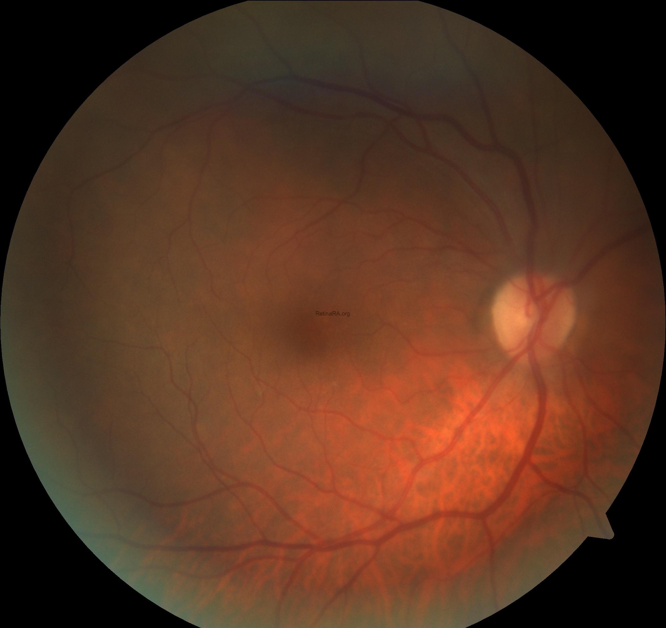
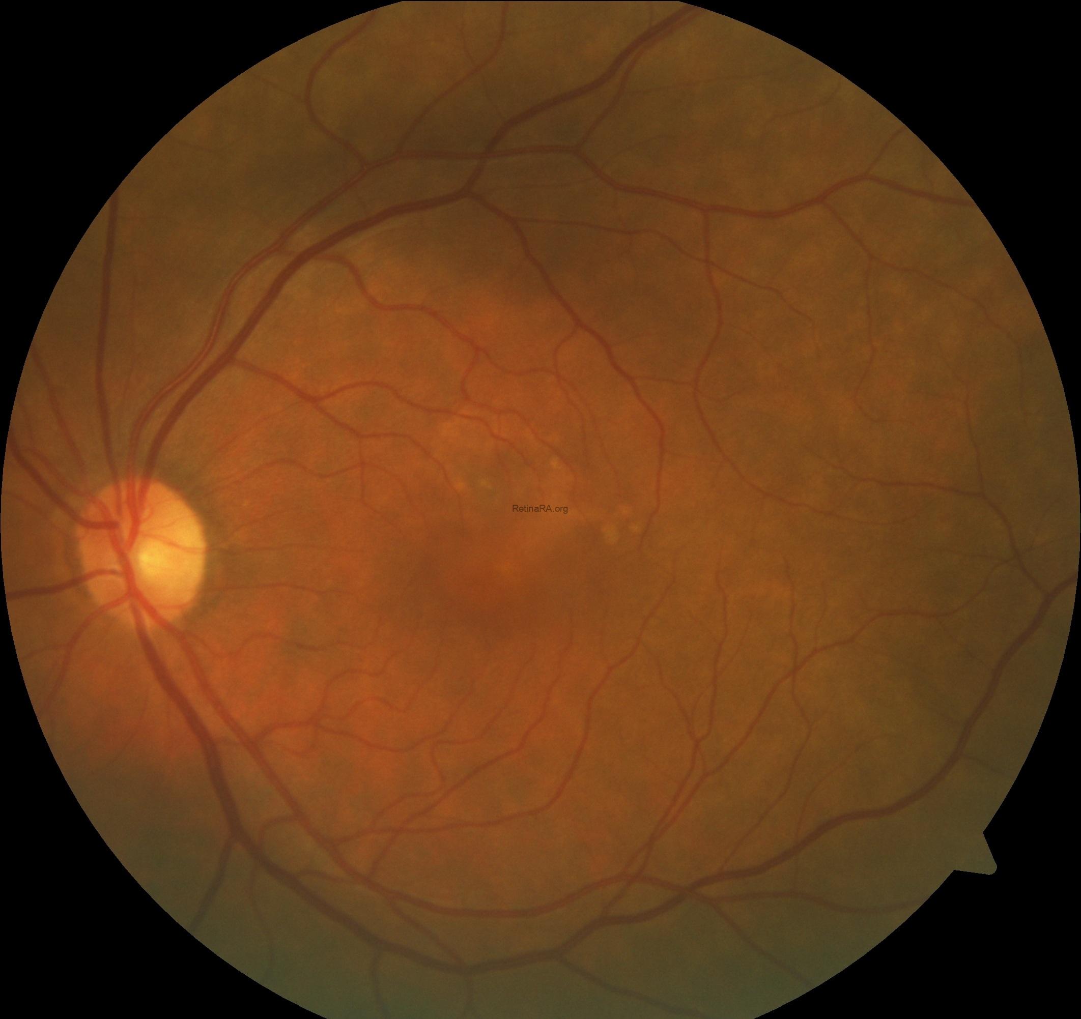
Color fundus photography revealed a normal-appearing posterior pole in the right eye. In the left eye, subtle RPE alterations and yellowish deposits were observed in the macular region, without obvious hemorrhage or extensive drusen.
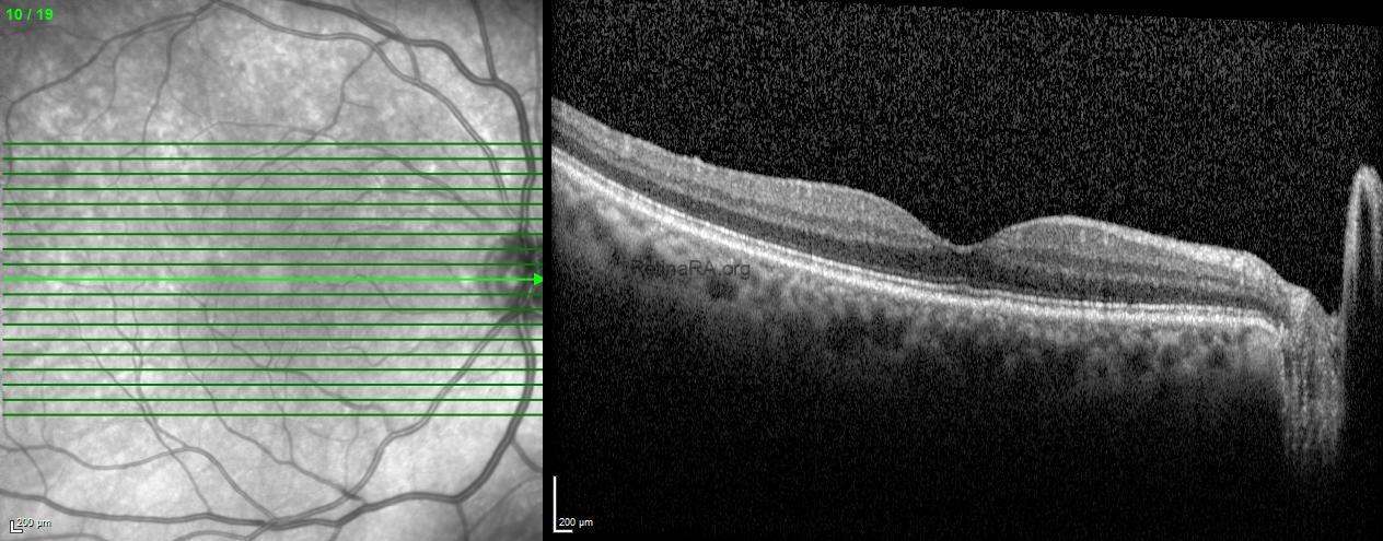
Spectral-domain OCT of the right eye demonstrated a preserved foveal contour and normal retinal architecture, without intraretinal or subretinal fluid.
In the left eye, OCT revealed an irregular pigment epithelial detachment associated with subretinal fluid and localized disruption of the outer retinal architecture. The irregular elevation of the RPE and underlying heterogeneous reflectivity raised suspicion for type 1 macular neovascularization .
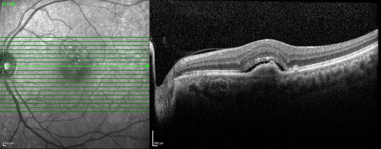
Enhanced depth imaging OCT demonstrated increased choroidal thickness with prominent dilated vessels in the outer choroid (pachyvessels), supporting an underlying pachychoroid phenotype.
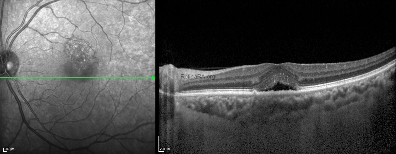
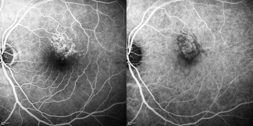
FFA of the left eye demonstrated an irregular area of macular hyperfluorescence corresponding to the RPE abnormalities and PED observed on OCT. Progressive hyperfluorescence and leakage were consistent with an active neovascular lesion. ICGA demonstrated an abnormal choroidal neovascular complex corresponding to the macular lesion.

En face OCTA revealed an abnormal vascular network corresponding to the PED, consistent with type 1 MNV. Cross-sectional OCTA with flow overlay demonstrated flow signals within the sub-RPE compartment, confirming the presence and anatomical localization of the neovascular complex.
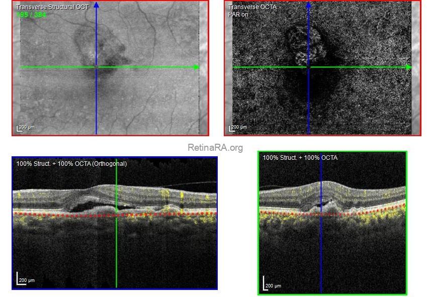
The combination of multimodal imaging findings established the presence of an active type 1 MNV associated with a pachychoroid phenotype.
Pachychoroid neovasculopathy (PNV) is characterized by type 1 macular neovascularization occurring in association with pachychoroid-related choroidal abnormalities, typically in the absence of conventional soft-drusen-associated age-related macular degeneration. Characteristic features include dilated outer choroidal vessels (pachyvessels), attenuation of the overlying inner choroidal layers, RPE alterations, and frequently increased choroidal thickness. Importantly, increased choroidal thickness alone is neither necessary nor sufficient for diagnosis, as the distribution and morphology of choroidal vessels are central to identifying the pachychoroid phenotype.
Differentiating PNV from chronic central serous chorioretinopathy without neovascularization and neovascular AMD is clinically important. Shallow or irregular PEDs and subretinal fluid may be present in both CSC and PNV; however, visualization of a neovascular network on OCTA provides direct evidence of MNV. In contrast to conventional neovascular AMD, PNV typically occurs in relatively younger patients and is associated with pachychoroid features rather than extensive soft drusen. ICGA further contributes to the evaluation of the choroidal vasculature and the characterization of neovascular lesions.
Credit: Kemal Tekin, M.D., FEBOS-R, from Ulucanlar Eye Training and Research Hospital
Instagram accounts: @retina.academy and @dr.kemaltekin
