

When vision loss tells a bigger story
A 58-year-old man presents directly to our ophthalmic emergency service with gradually worsening vision in his left eye over the past two weeks, accompanied by nasal visual field loss in his right eye. His presenting symptoms are exclusively ocular, with no systemic complaints.
Visual acuity:
• Right eye: 20/20
• Left eye: Hand movements
He reports no photopsia. His medical history is unremarkable apart from asthma.
Fundus examination reveals choroidal masses in both eyes, with macular involvement in the left eye.


B-scan ultrasonography demonstrates broad-based, dome-shaped choroidal masses in both eyes. A-scan shows medium-to-high internal reflectivity within the lesions, supporting the suspicion of bilateral choroidal metastases.
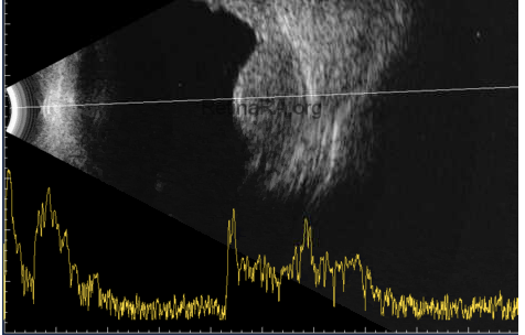
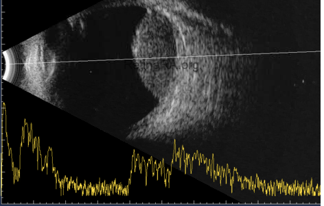
Chest radiography reveals hilar enlargement, prompting further staging with CT of the chest and abdomen and brain MRI. The patient is referred to the Department of Pulmonology for further evaluation and treatment.

The workup identifies findings consistent with metastatic lung cancer. Brain MRI demonstrates bilateral intraocular masses and falx-based soft-tissue lesions adjacent to the superior sagittal sinus in the high parietal region, more pronounced on the right than the left, with suspected sinus invasion.
Choroidal metastases may be the first sign of an underlying malignancy, even in patients without systemic symptoms. This case highlights how recognizing bilateral choroidal masses and initiating prompt systemic investigation can uncover a previously undiagnosed cancer.
Credit: Houssein El Hajjami
Department of Ophthalmology, Klinikum Kassel, Kassel, Germany
Instagram account: @dr.elhajjami
