

A 9-year-old boy was referred for evaluation of bilateral decreased visual acuity. He had no known systemic disease. Best-corrected visual acuity was 20/32 in the right eye and 20/40 in the left eye. Intraocular pressures were within normal limits, and anterior segment examination was unremarkable in both eyes.
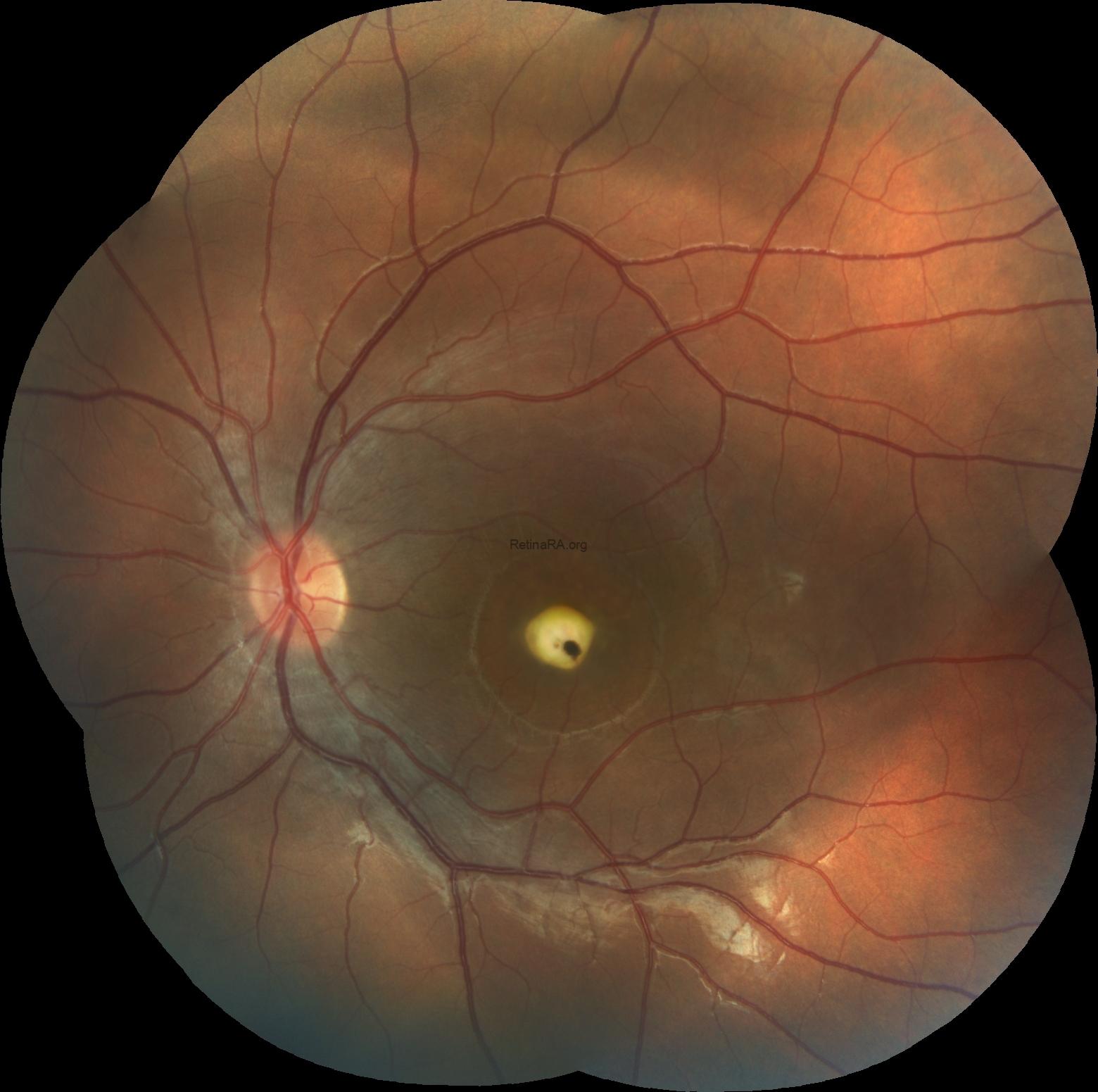
Color fundus photography demonstrated multiple well-circumscribed yellowish subretinal deposits clustered within the macula of the right eye. The left eye showed an elevated yellow-white subfoveal lesion with focal central hyperpigmentation and surrounding smaller yellowish deposits, giving a fibrotic-appearing configuration. Widefield imaging confirmed that the abnormalities were confined to the posterior pole, with no evident involvement of the midperipheral or peripheral retina in either eye


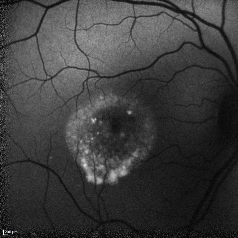
Fundus autofluorescence revealed heterogeneous, predominantly increased autofluorescence corresponding to the macular deposits in the right eye. In the left eye, a ring of increased autofluorescence surrounded a central area of reduced autofluorescence, reflecting heterogeneous accumulation of vitelliform material and central retinal pigment epithelium disturbance.

Spectral-domain optical coherence tomography of the right eye demonstrated a macular detachment with an optically hyporeflective subretinal space and irregular hyperreflective material along the photoreceptor–retinal pigment epithelium interface. In the left eye, OCT showed a markedly elevated hyperreflective subfoveal lesion with posterior shadowing and surrounding hyporeflective subretinal spaces, consistent with advanced fibrotic change.

The multimodal imaging findings were consistent with Best vitelliform macular dystrophy, with marked interocular asymmetry and different stages of disease progression between the two eyes. Electro-oculography demonstrated an absent light rise, indicating severe dysfunction of the retinal pigment epithelium and providing additional support for the diagnosis. Molecular genetic testing for BEST1 was recommended for diagnostic confirmation and genetic counselling.
Best vitelliform macular dystrophy is an inherited retinal pigment epithelium disorder, most commonly caused by autosomal dominant variants in the BEST1 gene. It typically presents in childhood with bilateral, although often asymmetric, yellowish vitelliform deposits at the macula. The lesions may progress through vitelliform, pseudohypopyon, vitelliruptive, atrophic, and fibrotic stages, and different stages may coexist between the two eyes. Fundus autofluorescence and OCT are useful for characterizing the accumulated material and associated structural changes. Electro-oculography classically demonstrates a reduced or absent light rise, reflecting impaired retinal pigment epithelium function. Patients should be monitored for progressive atrophy, fibrosis, and secondary choroidal neovascularization.
Credit: Kemal Tekin, M.D., from Ulucanlar Eye Training and Research Hospital
Instagram accounts: @retina.academy and @dr.kemaltekin
