

A patient with a history of inactive thyroid orbitopathy was evaluated using multimodal retinal imaging. Colour fundus photography demonstrated bilateral horizontal-to-oblique chorioretinal folds involving the posterior pole, extending from the peripapillary region toward the macula. Tilted optic discs and peripapillary atrophy were also noted.
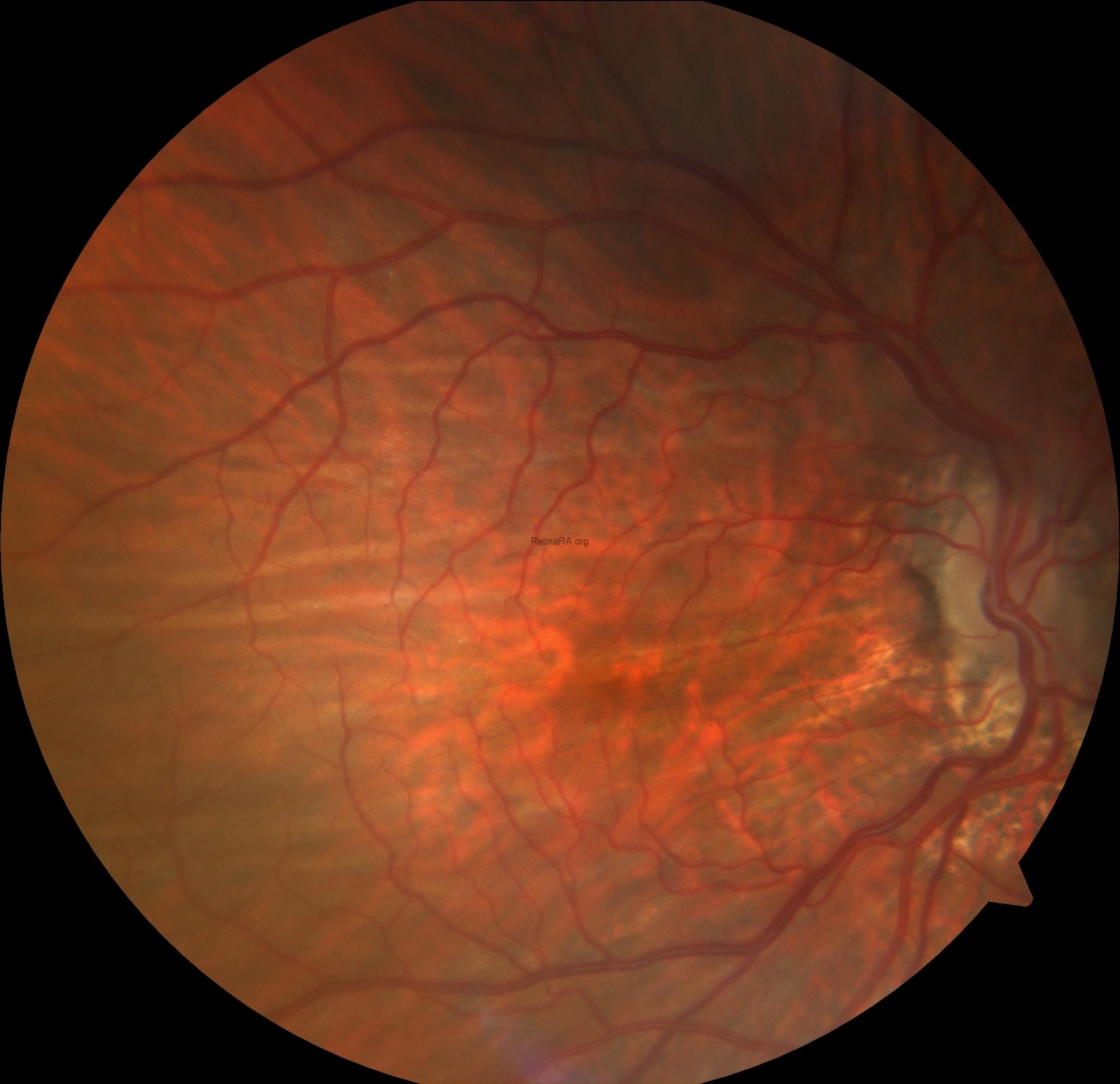
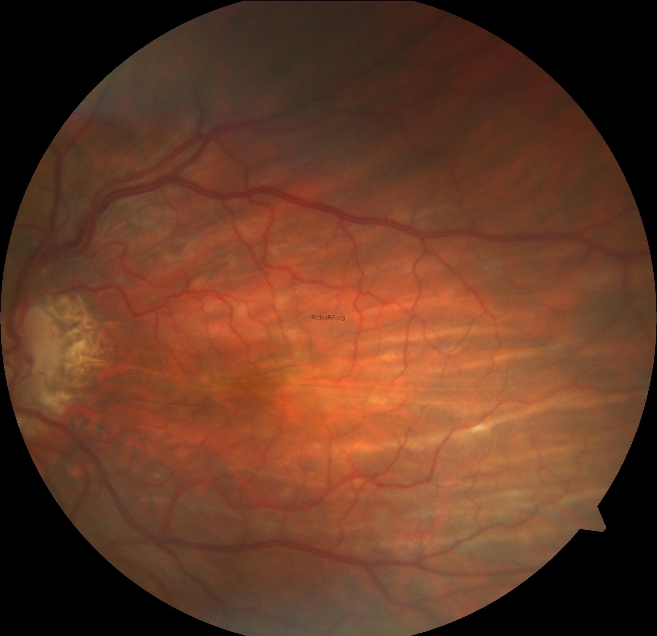
Multicolour imaging enhanced the visibility of the folds, showing alternating bright and dark curvilinear lines across the posterior pole.


Fundus autofluorescence revealed corresponding alternating hyperautofluorescent and hypoautofluorescent streaks.


Spectral-domain optical coherence tomography through the folds demonstrated characteristic undulations of the retinal pigment epithelium–Bruch’s membrane complex and underlying choroid, while the inner retinal surface remained relatively smooth and retinal lamination was preserved.

Choroidal folds represent parallel undulations of the RPE–Bruch’s membrane–choroid complex. They may be observed in several conditions, including hyperopia, ocular hypotony, papilloedema, orbital masses, and thyroid orbitopathy. In thyroid orbitopathy, orbital congestion, extraocular muscle enlargement, and posterior globe compression may contribute to fold formation. Importantly, choroidal folds may persist even during the inactive phase of thyroid orbitopathy. Recognition of this association is clinically relevant, as multimodal imaging can help distinguish chronic structural folds from active inflammatory or exudative retinal disease.
Credit: Kemal Tekin, M.D., from Ulucanlar Eye Training and Research Hospital
Instagram accounts: @retina.academy and @dr.kemaltekin
