

A 20-year-old woman presented with progressive bilateral central visual loss, impaired color vision, and prominent photophobia. The BCVA was 20/200 in both eyes. Anterior segment examination was unremarkable bilaterally.
Multicolor fundus imaging demonstrated bilateral central macular pigmentary alteration with a relatively symmetric appearance. No marked peripheral retinal degeneration, vascular attenuation, or optic disc pallor was evident.


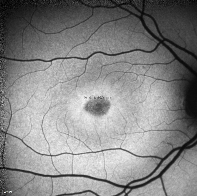
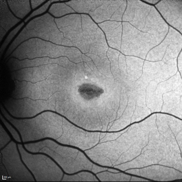
Fundus autofluorescence revealed a bilateral bull’s-eye maculopathy pattern, characterized by central hypoautofluorescence surrounded by a ring of relatively increased autofluorescence, consistent with macular photoreceptor–retinal pigment epithelium dysfunction.
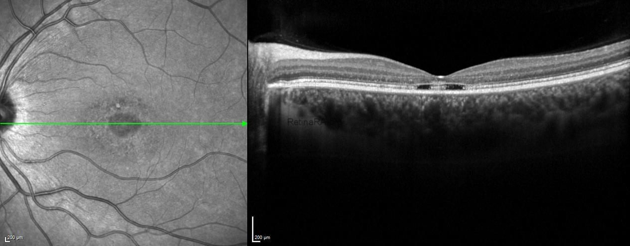
Spectral-domain optical coherence tomography showed bilateral foveal outer retinal disruption, including attenuation/loss of the ellipsoid zone and focal outer retinal cavitary changes, while the inner retinal architecture was relatively preserved.

Full-field electroretinography was consistent with cone dystrophy, showing markedly reduced photopic responses with relatively preserved scotopic responses.
Cone dystrophy is an inherited retinal disorder primarily affecting cone photoreceptors. Patients typically present in childhood or early adulthood with reduced central visual acuity, impaired color vision, and severe photophobia. Fundus findings may be subtle in early stages, making multimodal imaging and electrophysiologic testing essential for diagnosis. Genetic testing is recommended to confirm the diagnosis, determine the inheritance pattern, guide family counseling, and identify potential eligibility for future therapeutic trials. Regular follow-up with multimodal imaging is useful for monitoring structural progression and visual prognosis.
Credit: Kemal Tekin, M.D., from Ulucanlar Eye Training and Research Hospital
Instagram accounts: @retina.academy and @dr.kemaltekin
