

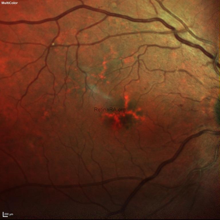

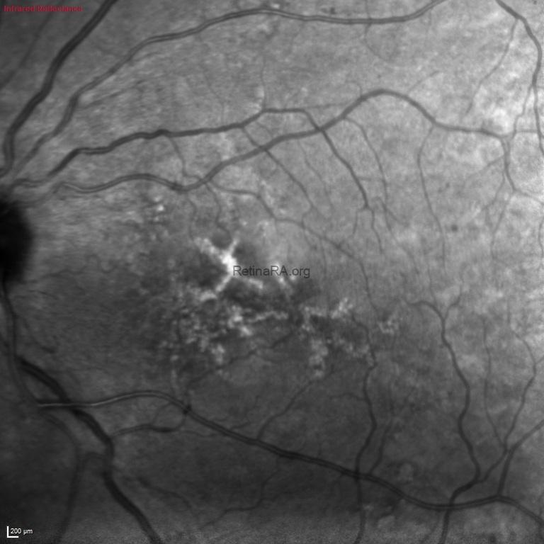
Infrared reflectance imaging highlighted the macular pigmentary deposits more clearly, showing irregular hyperreflective and hyporeflective changes corresponding to the patterned retinal pigment epithelium alterations.

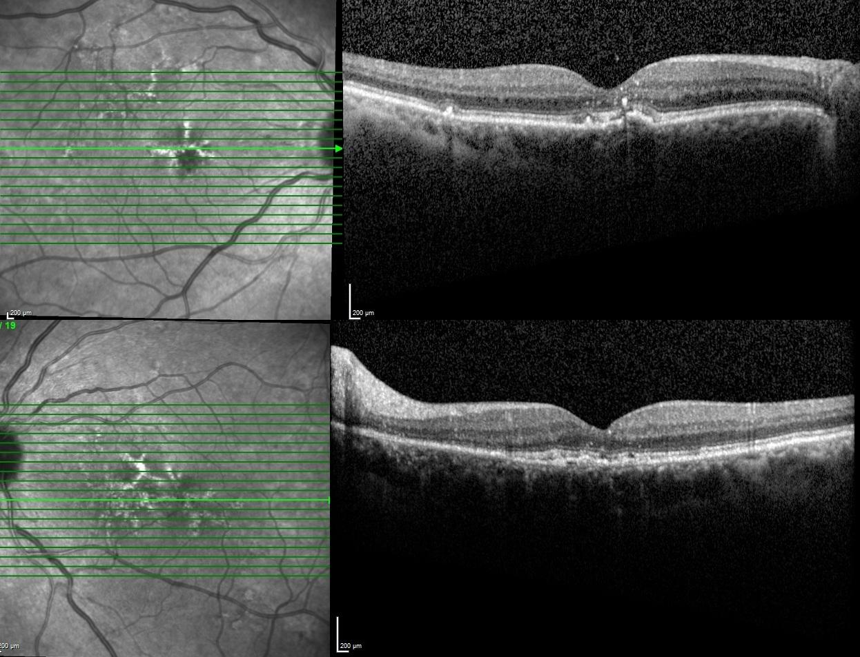
Spectral-domain optical coherence tomography revealed focal hyperreflective material at the level of the retinal pigment epithelium/photoreceptor complex, with mild disruption of the outer retinal bands in both eye. The foveal contour was relatively preserved, and there was no intraretinal or subretinal fluid.
Butterfly-shaped pattern dystrophy is a subtype of pattern dystrophy of the retinal pigment epithelium, typically presenting in middle-aged or older adults. It is characterized by bilateral, often symmetric pigment deposition at the macula in a butterfly-like pattern. The condition is usually slowly progressive and may remain visually mild for many years.
Multimodal imaging is useful for distinguishing butterfly-shaped pattern dystrophy from age-related macular degeneration and other macular disorders. OCT is particularly important for detecting outer retinal disruption, monitoring structural progression, and identifying complications such as choroidal neovascularization.
Genetic testing may be considered, especially in familial cases, as pattern dystrophies may be associated with mutations in genes such as PRPH2/RDS. Long-term follow-up is recommended to monitor visual function and possible secondary neovascular complications.
Credit: Kemal Tekin, M.D., from Ulucanlar Eye Training and Research Hospital
Instagram accounts: @retina.academy and @dr.kemaltekin
