Transient superficial retinal infiltrates one of the most characteristic features of Behçet’s uveitis and even one of them is considered as posterior segment involvement. The use of OCT has provided insight into the nature of these lesions. SD OCT sections passing through retinal infiltrates typically show focal retinal thickening, increased hyper-reflectivity with blurring of inner retinal layers, and optical shadowing. However unlike a focus of retinochoroiditis, such as in ocular toxoplasmosis, there is no focal choroidal thickening beneath the retinal infiltrate and the RPE contour is unaffected, that is, there is no inward bowing of the RPE line.


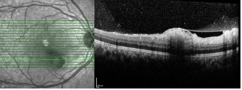
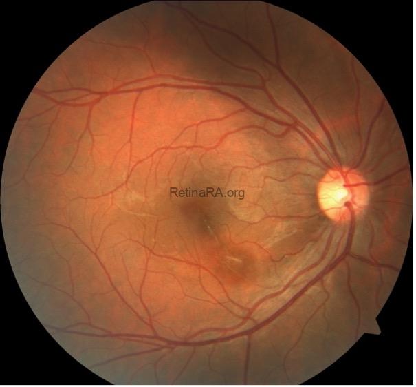
Three months later, color fundus photography shows a focal retinal nerve fiber layer defect as a sequel of the infiltrate and the optical coherence tomography shows focal disorganization and hyper-reflectivity in the inner retina and loss of the retinal nerve fiber layer. In Behçet uveitis, retinal infiltrates rapidly resolve without any significant retinochoroidal scarring. However, SD OCT sections typically show inner retinal atrophy.

Credit: Merve İnanç Tekin, M.D., from Ulucanlar Eye Training and Research Hospital
Instagram accounts: @uveacademy and @merveinanctekin
