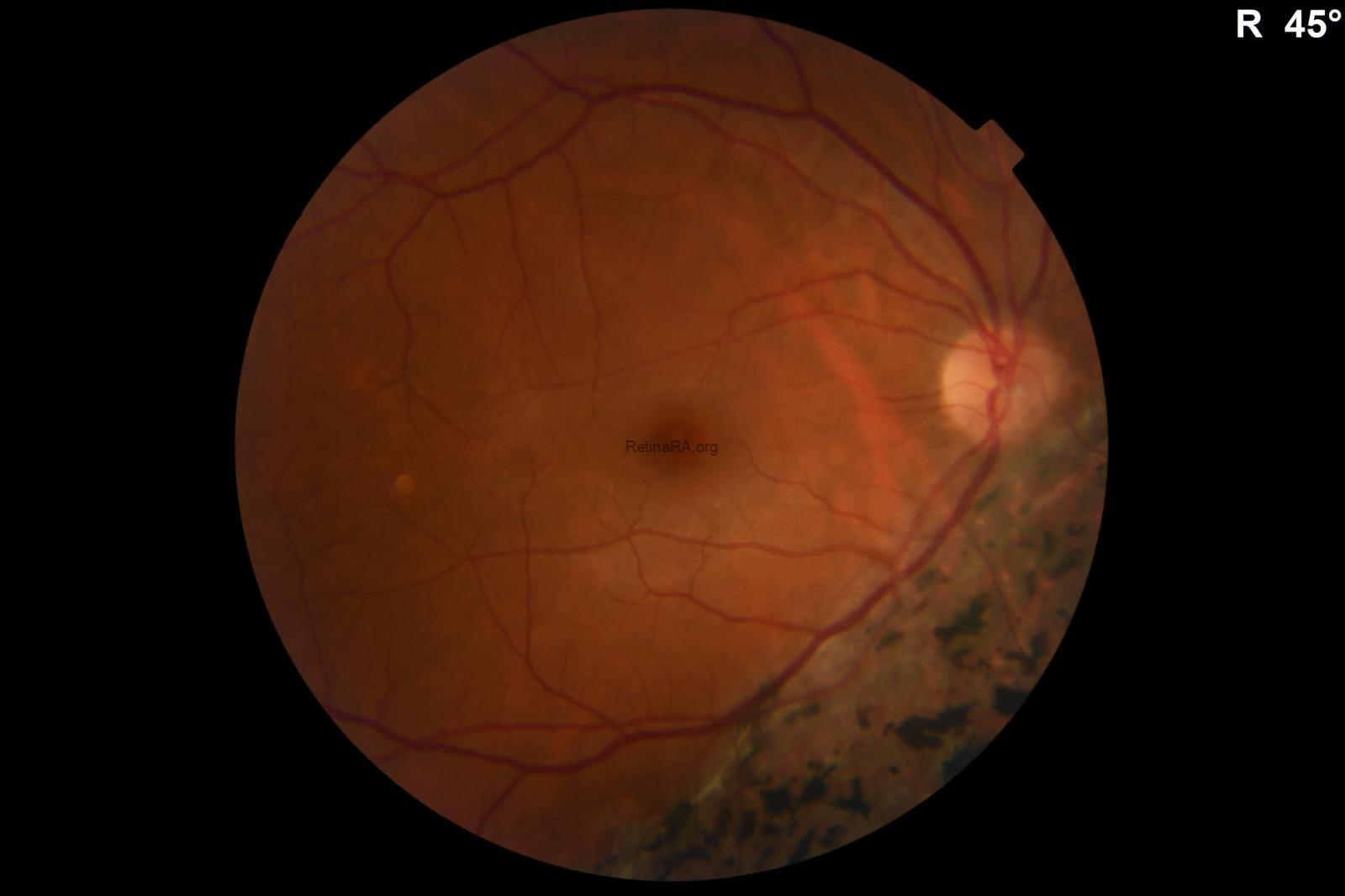
A 25-year-old female patient presented for refractive surgery evaluation. Best-corrected visual acuity was 20/20 in both eyes. Anterior segment examination was completely unremarkable bilaterally. Fundus examination of the left eye was entirely normal.
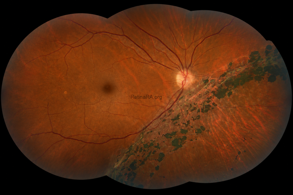
Fundus examination of the right eye revealed extensive pigmentary alterations and retinal pigment epithelium (RPE) atrophy predominantly located inferior to the optic disc, extending toward the inferior peripheral retina. The lesion initially raised suspicion for segmental retinitis pigmentosa due to the prominent bone-spicule–like pigment accumulation.
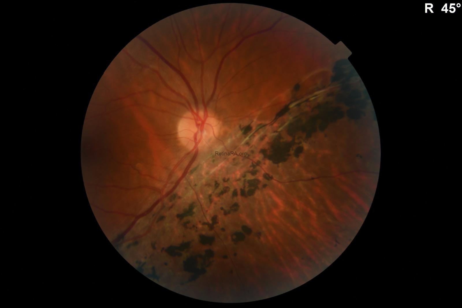
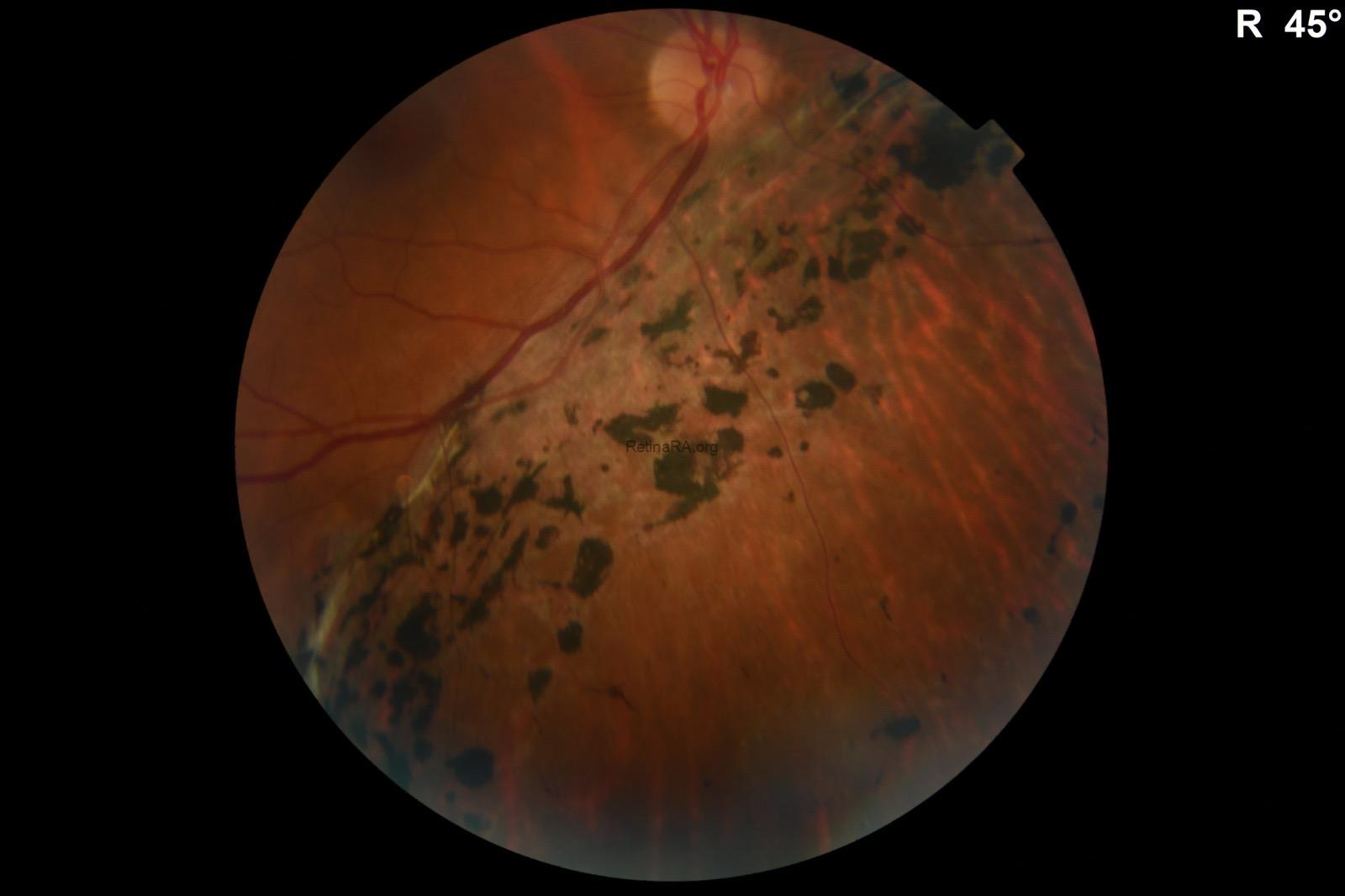
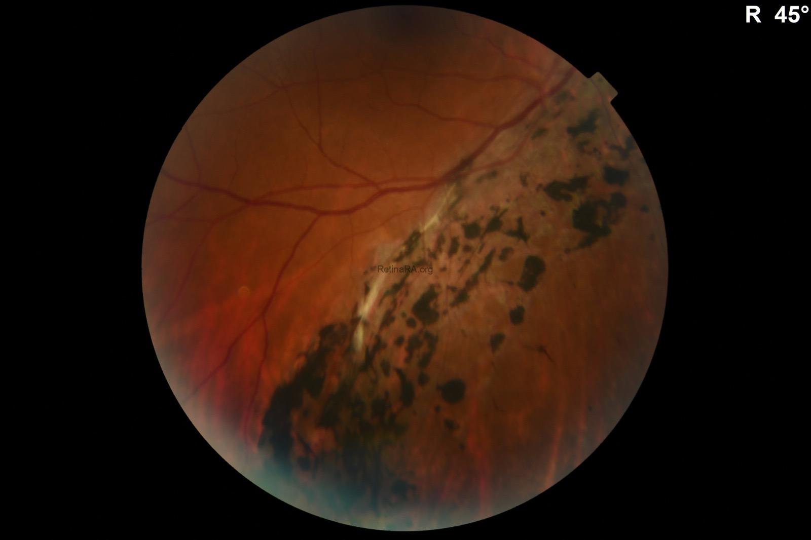
However, several clinical findings favored a diagnosis of traumatic pigmentary retinopathy rather than segmental retinitis pigmentosa. First, the pigmentary changes did not conform to a single retinal segment or a typical sectoral distribution pattern expected in segmental RP. Instead, the lesions demonstrated an irregular and more diffuse inferior extension.
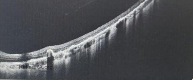
In addition, linear crack-like structures were observed between the pigmentary areas. These linear lesions appeared deeper than the level of the retinal pigment epithelium and were thought to possibly originate from Bruch’s membrane and/or the choroid, suggesting previous traumatic damage rather than a primary inherited retinal dystrophy.
Detailed history taking revealed a history of blunt ocular trauma at the age of 4 years, as reported by the patient’s mother. This history further supported the diagnosis of traumatic pigmentary retinopathy.
To exclude retinitis pigmentosa, full-field electroretinography (ERG) was performed and demonstrated normal retinal function in both eyes. The normal ERG findings were considered inconsistent with retinitis pigmentosa and supportive of a localized traumatic process.
Overall, the combination of unilateral involvement, normal fellow eye findings, atypical distribution of pigmentary changes, presence of linear traumatic-appearing cracks, preserved visual acuity, and normal ERG findings supported the diagnosis of traumatic pigmentary retinopathy mimicking segmental retinitis pigmentosa.
Credit:
Case: Gunel Agayeva, Md
Badam Medical Center, Baku, Azerbaijan
Instagram account: @dr.gunel_agayeva_phd
Caption: M. Giray Ersoz, MD, FEBO
Memorial Hospital, Department of Ophthalmology, Istanbul, Turkey
Arel University School of Medicine, Department of Ophthalmology, Istanbul, Turkey
Instagram accounts: @retina.review and @retina.dr.girayersoz
