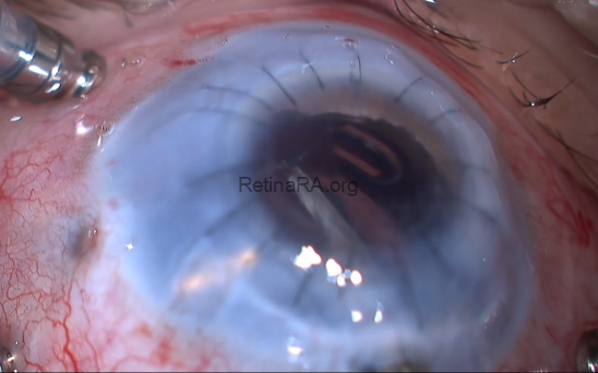
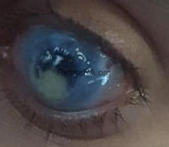
A 15-year-old male patient with vision loss and pain in the left eye for the last 5 years. He had a history of bilateral congenital glaucoma and congenital cataract surgery. Later, evisceration was performed on the right eye and a prosthesis was placed. His left eye was completely whitened and painful for the last 5 years. Buphthalmus and total corneal leukoma were present on the left eye examination. The anterior segment and fundus cannot be visualized. The day after penetrating keratoplasty was performed on the patient, a total crystalline lens and a 4-haptic IOL were seen in the patient’s vitreous. Pars plana vitrectomy was planned after the patient was followed up for 1 month and the crystalline lens material was seen to be in contact with the endothelium. Due to high IOP values despite topical treatment, endolaser cyclophotocoagulation was also planned. IOL implantation was not performed because the patient was buphthalmic and the axial length was too long.
Credit: M. Giray Ersoz, MD, FEBO
Biruni University School of Medicine, Department of Ophthalmology, Istanbul, Turkey
Instagram accounts: @retina.review and @retina.dr.girayersoz
