This was a 20-year-old healthy man without any systemic disease who admitted with an acute-onset visual field defect in his right eye. He also suffered from intermittent headaches.
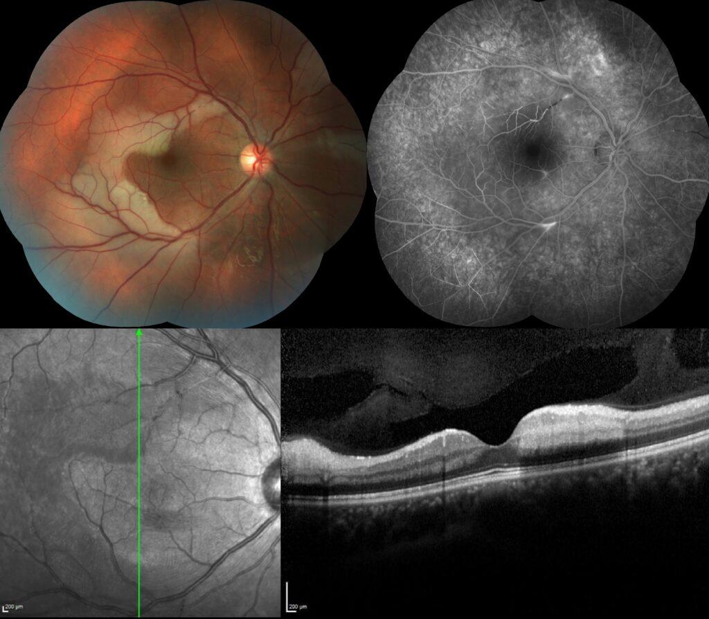
Fundus examination of the right eye showed crescent shaped retinal ischemic whitening involving the superior and inferior fovea with multiple retinal arterial wall plaques called as Gass plaques.

Fundus fluorescein angiography revealed the patched arterial wall hyper-fluorescence of Gass plaques in addition to obstruction of midarteriolar segment located at the superior of fovea.

Vertical spectral-domain optical coherence scan of the retinal ischemic areas demonstrated hyper-reflectivity of inner retinal layers with posterior shadowing.

In light of these multimodal imaging findings, the preliminary diagnosis was probable Susac syndrome and therefore, further medical examination was carried out by the Otorhinolaryngology and Neurology departments. While audiometry was normal; MRI imaging was reported as white matter disturbances affecting peri-corpus callosum compatible with Susac syndrome.
Susac syndrome is a rare autoimmune mediated brain–eye–ear syndrome affecting the small arterial vessels of the brain, inner ear and retina. It is characterized by the clinical triad of encephalopathy, branch retinal artery occlusions and sensorineural hearing loss, usually affecting low‐ to mid‐frequencies. The classic triad of features is present in only 13% of patients at onset, yet over 80% of patients eventually develop all symptoms.
Credit: Kemal Tekin, M.D., from Ulucanlar Eye Training and Research Hospital
Instagram accounts: @retina.academy and @dr.kemaltekin
