
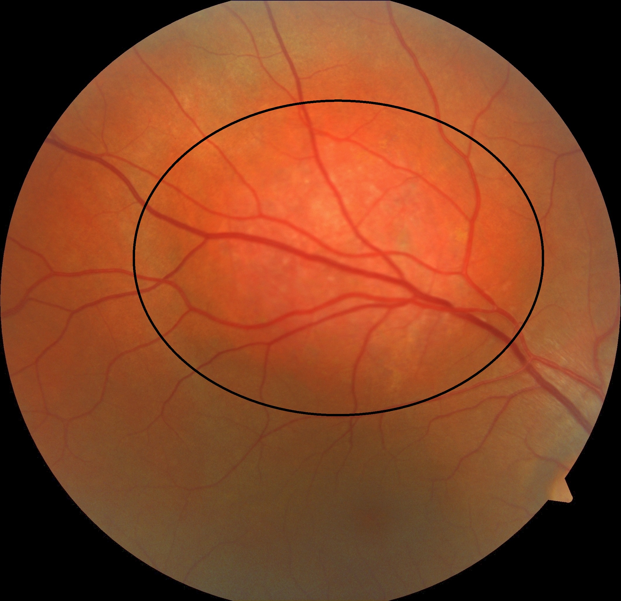
A 48-year-old male with circumscribed choroidal hemangioma and accompanying subretinal fluid. The tumor appears as an orange-red elevated mass on color fundus photography and hypoautofluorescence on FAF. OCT shows dome-shaped smooth choroidal mass and subretinal fluid. FA demonstrates hyperfluorescence in the early and late stages. The lesion appears hyperfluorescence in the mid-stage ICG and very late-stage ICG reveals the wash-out phenomenon. One month after full-dose PDT with intravenous bolus Visudyne injection, the subretinal fluid completely resolved and visual acuity increased from 20/60 to 20/20.
For the treatment of choroidal hemangioma, either standard treatment settings, a bolus protocol, or a high-fluence protocol can be used. A bolus protocol is thought to reduce washout. ‘Bolus’ protocol: verteporfin is rapidly infused (bolus) over 1 minute; PDT treatment begins exactly 6 minutes after the start of the verteporfin infusion or is infused within 4-5 minutes and PDT treatment begins immediately after the infusion. It is applied with 600 mW/cm2 radiation, 83 seconds and 50 J/m2.
Credit: M. Giray Ersoz, MD, FEBO
Biruni University School of Medicine, Department of Ophthalmology, Istanbul, Turkey
Instagram accounts: @retina.review and @retina.dr.girayersoz
Color fundus photography of circumscribed choroidal hemangioma

Fundus autofluorescence imaging of circumscribed choroidal hemangioma

Early-stage fluorescein angiography of circumscribed choroidal hemangioma

Late-stage fluorescein angiography of circumscribed choroidal hemangioma

Mid-stage indocyanine green angiography of circumscribed choroidal hemangioma

Very late-stage indocyanine green angiography of circumscribed choroidal hemangioma (the wash-out phenomenon)

Pre-PDT optic coherence tomography scans

Post-PDT optic coherence tomography scans

