This was a 15-year-old girl admitted for routine eye check-up. The best corrected visual acuities were 20/20 for both eyes and anterior segments were unremarkable.
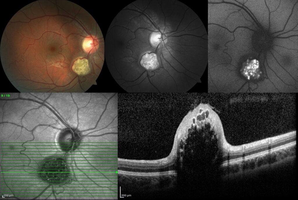
Fundus examination of the right eye showed a yellowish, opaque, well-circumscribed, elevated solid lesion clustered spherules formed a mulberry-like shape inferior to the optic disc; while the left eye was completely normal.


The lesion borders and its relationship with the vessels were more clearly visible in the red-free image.

Fundus autofluorescence demonstrated hyperautofluorescent cystic-like opacities and calcifications giving a mulberry-like appearance.

Spectral-domain optical coherence tomography exhibited dome-shaped thickening of inner retinal layers, multiple intraretinal moth-eaten optically empty cystic spaces with posterior shadowing.

In light of these multimodal imaging findings, the diagnosis was retinal astrocytoma and further medical examination was carried out by dermatology and neurology departments for the association of other systemic diseases, and the patient was diagnosed as tuberous sclerosis complex.
Retinal astrocytoma (RA), also known as retinal astrocytic hamartoma, is a rare tumour arising from retinal glial cells. Most patients with RA are asymptomatic, thus RA is usually discovered as an incidental finding. RA is diagnosed clinically based on its characteristic ophthalmoscopic appearance and it is the best-known ocular manifestation of tuberous sclerosis. However, it can also be idiopathic or associated with neurofibromatosis.
Credit: Kemal Tekin, M.D., from Ulucanlar Eye Training and Research Hospital
Instagram accounts: @retina.academy and @dr.kemaltekin
