A 28-year-old female patient was referred for evaluation of blurred optic disk margins in both eyes. The patient was completely healthy without any systemic disease. Her ocular history was also unremarkable. The BCVAs were 20/25 for both eyes and IOPs were within normal limits. Pupils were round and reactive to light without any afferent pupillary defects. Slit lamp biomicroscopy was also unremarkable for both eyes.
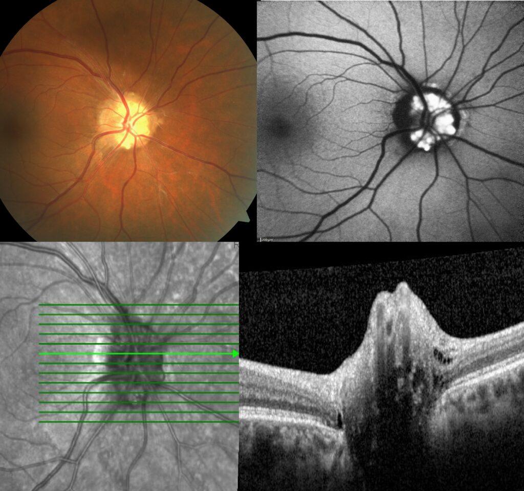
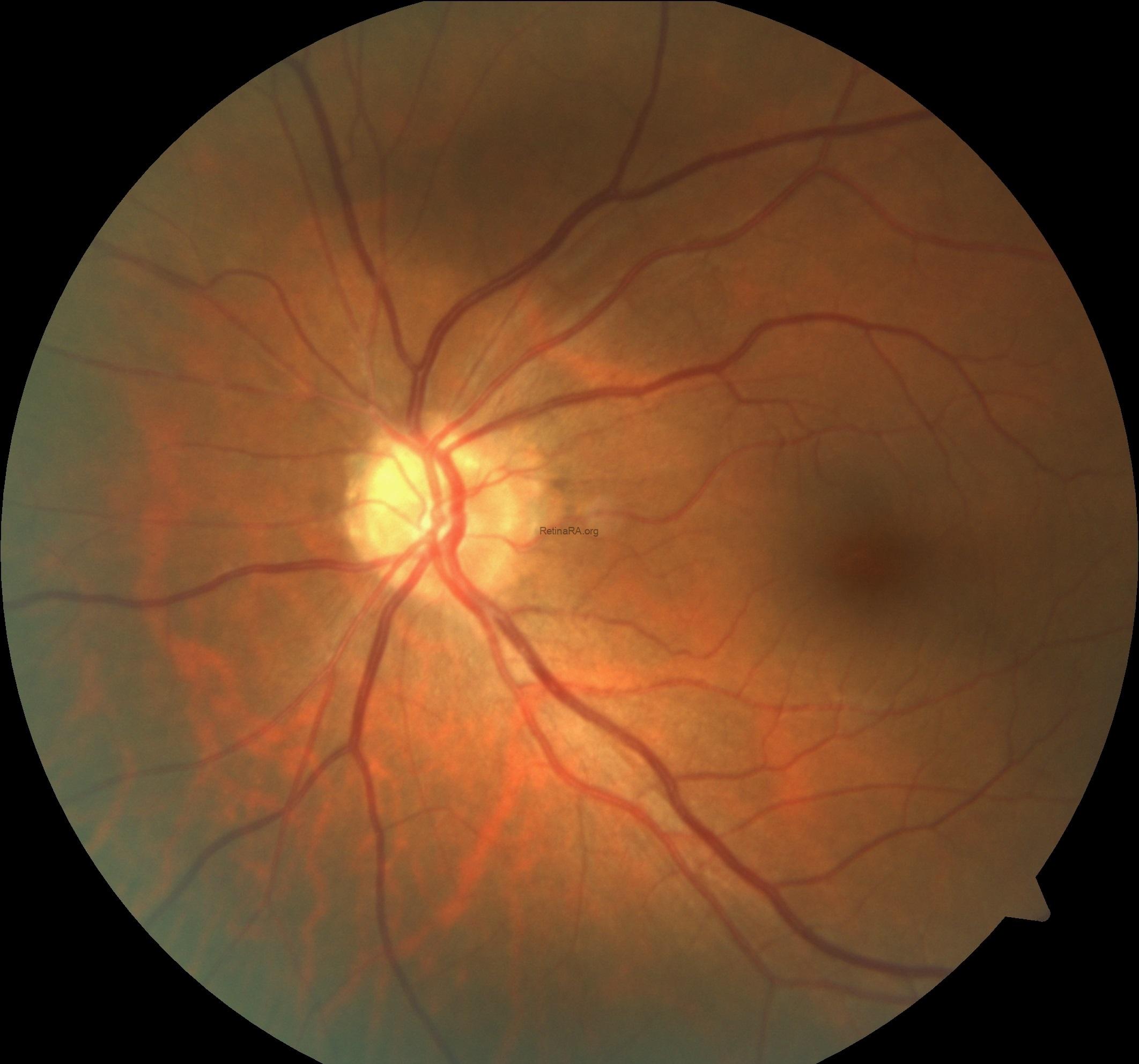
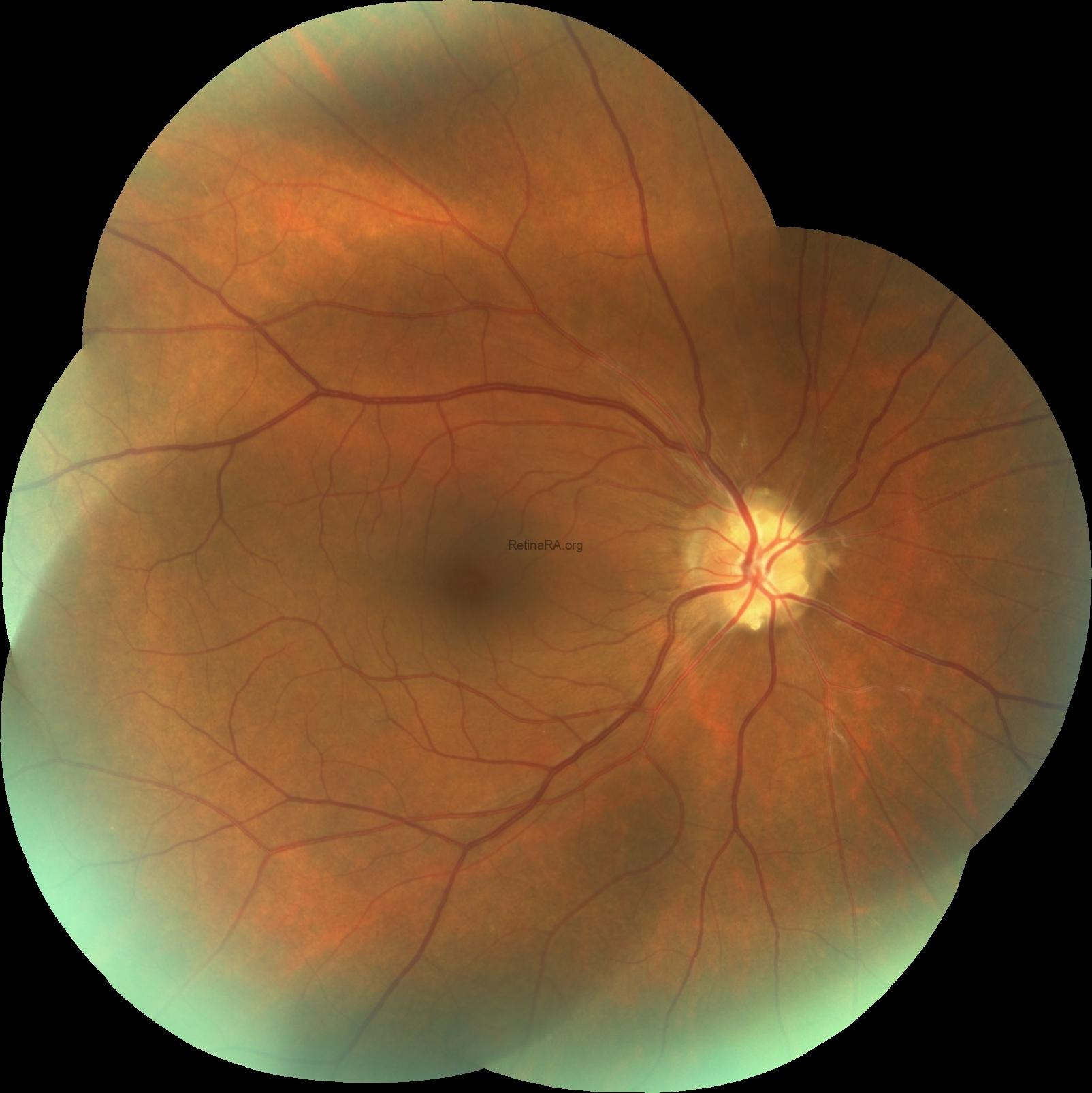
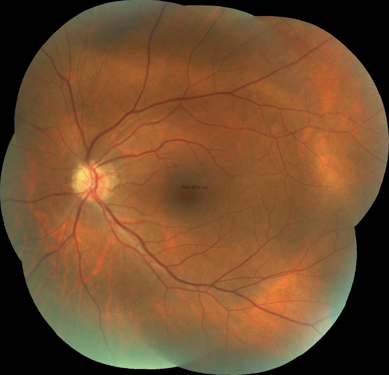
Fundus examination showed a scalloped and raised optic nerve giving the appearance of indistinct and irregular disc margins in both eyes. This was more evident in the right eye and yellowish-white nodules were also detected in the right eye. The macula, vessels and peripheral retina were normal for both eyes.

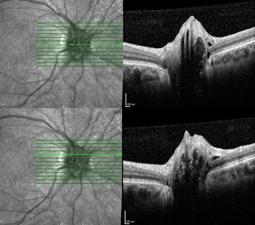
Optical coherence tomography scans passing through optic nerve demonstrated dome-shaped elevation of the optic nerve head with hyporeflective amorphous internal cores surrounded by hyperreflective borders.

Fundus autofluorescence imaging exhibited brightly autofluorescing within the optic disc of both eyes.


Based on multimodal imaging findings, the diagnosis was optic nerve head drusen. The patient was advised to undergo regular follow-up without any treatment or intervention.
Optic nerve head drusen (ONHD) are acellular, calcified deposits located within the optic nerve head. They can mimic papilledema on fundus examination, making accurate diagnosis essential. Multimodal imaging plays a crucial role: optical coherence tomography often reveals a lumpy, irregular optic disc contour, while B-scan ultrasonography and fundus autofluorescence can confirm the presence of drusen. Although most patients remain asymptomatic, ONHD may be associated with visual field defects due to axonal damage. No specific treatment is required; instead, regular follow-up is recommended to monitor for progressive changes. Early recognition of ONHD prevents unnecessary interventions for suspected papilledema and ensures appropriate long-term management.
Credit: Kemal Tekin, M.D., from Ulucanlar Eye Training and Research Hospital
Instagram accounts: @retina.academy and @dr.kemaltekin
