
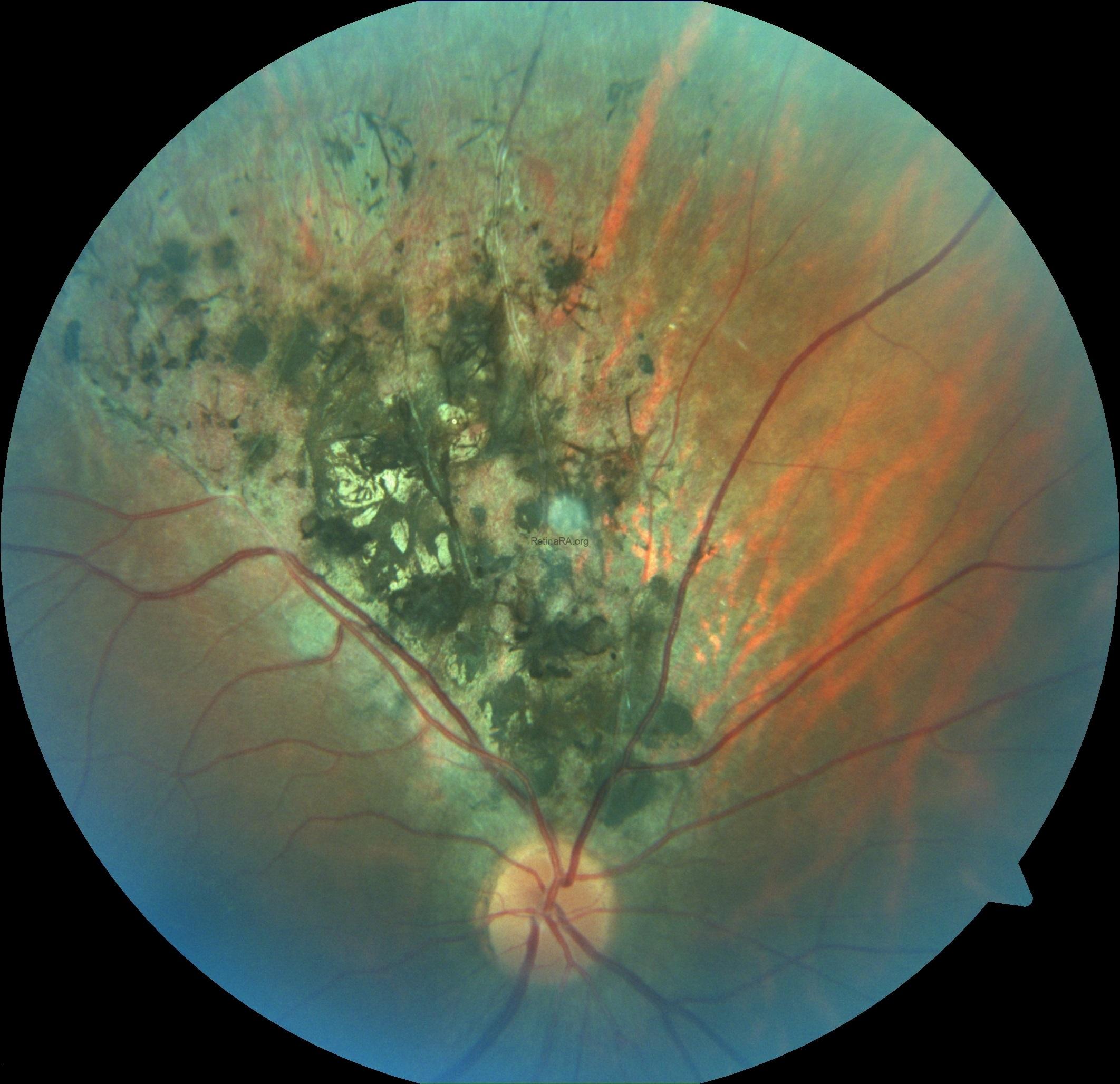
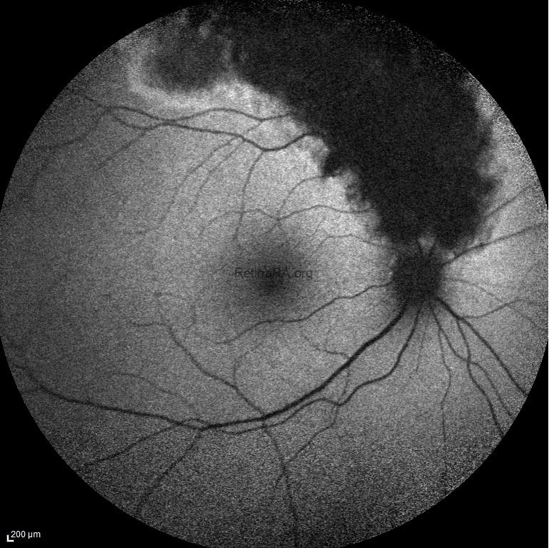
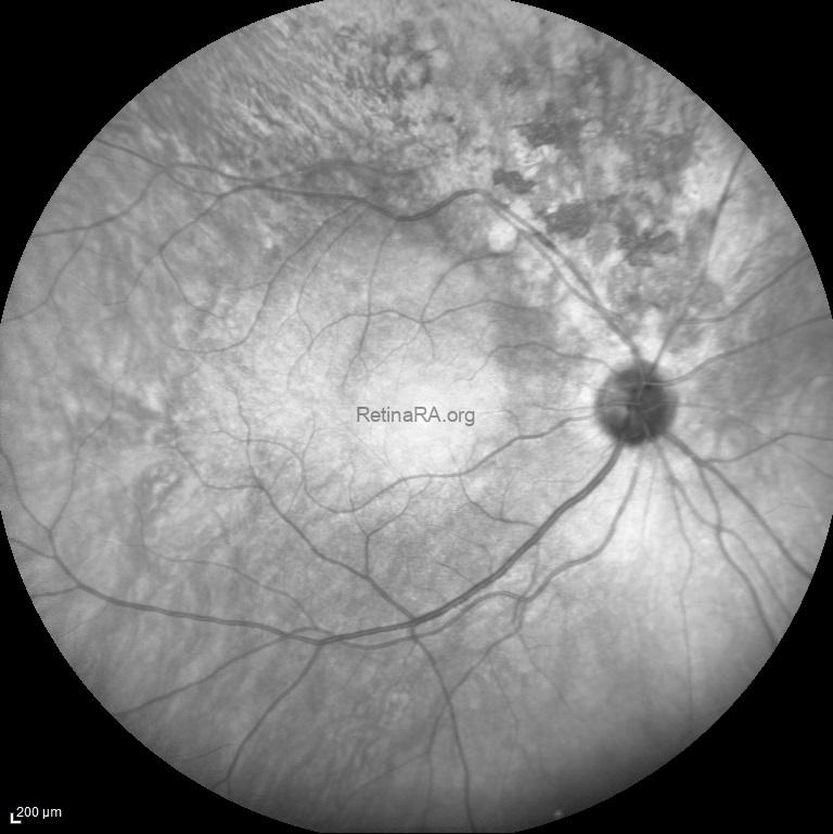
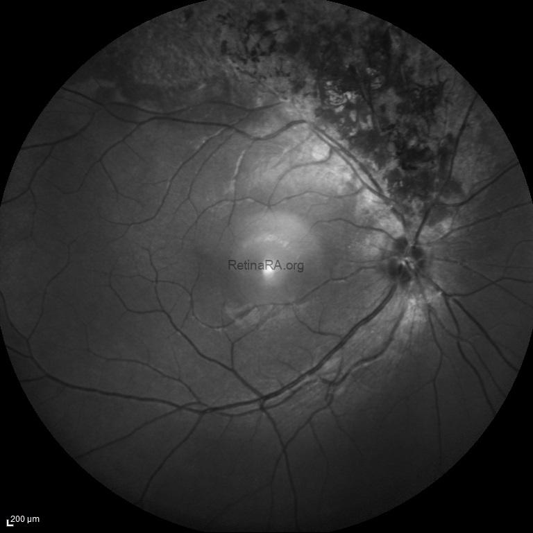
Traumatic pigmentary retinopathy was detected in a 38-year-old male with a history of blunt eye trauma and hyphema. The ophthalmic evaluation of the left eye was normal. His visual acuity was 20/20 bilaterally. Color fundus photograph, fundus autofluorescence image, infrared reflectance image, and blue reflectance image of the right eye show pigmentary changes at the anterior of the superior-temporal arcade simulating “sector retinitis pigmentosa”.
Color fundus photography of traumatic pigmentary retinopathy
Fundus autofluorescence of traumatic pigmentary retinopathy
Infrared reflectance image of traumatic pigmentary retinopathy
Blue reflectance image of traumatic pigmentary retinopathy
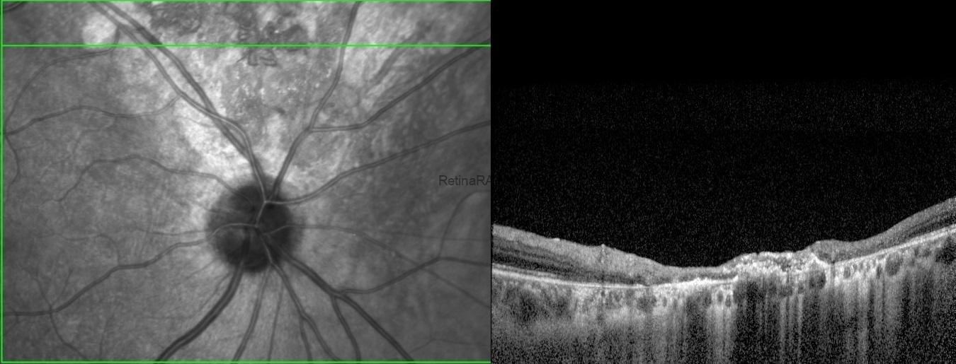
Severe outer and inner retinal deterioration is seen on OCT. However, it does not look like the classic retinitis pigmentosa appearance (look – https://www.retinara.org/category/retinitis-pigmentosa/ ). It is important to make the differential diagnosis of traumatic pigmentary retinopathy with retinitis pigmentosa and autoimmune retinopathies.
Credit: M. Giray Ersoz, MD, FEBO
Biruni University School of Medicine, Department of Ophthalmology, Istanbul, Turkey
Instagram accounts: @retina.review and @retina.dr.girayersoz
