This was a 22-year-old male who was referred for photodynamic therapy due to bilateral central serous chorioretinopathy. It has been learnt that the patient’s father has also undergone photodynamic therapy and intravitreal injections before.
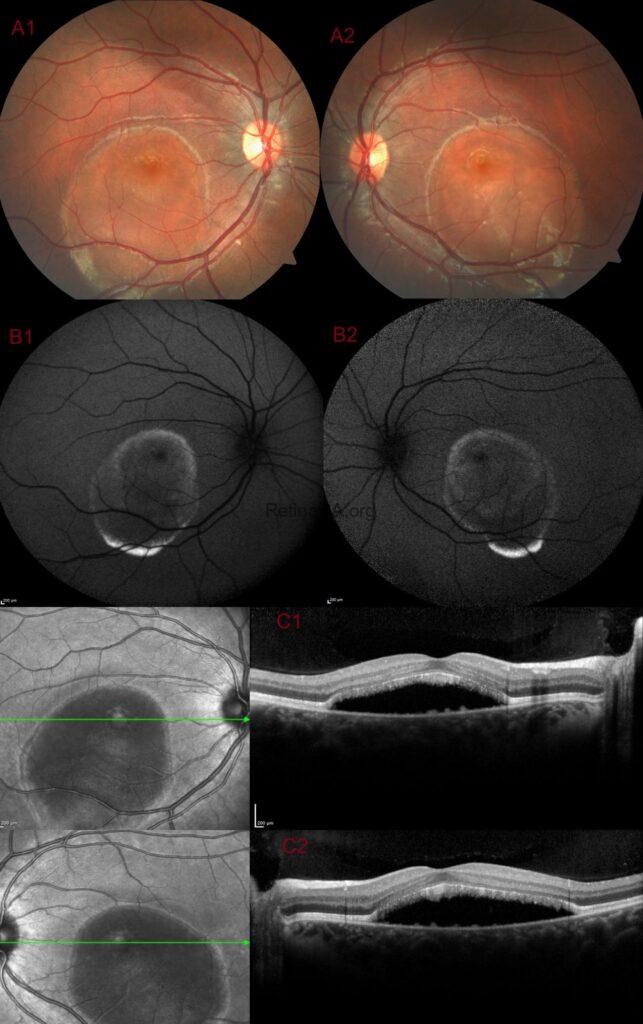
Fundus examinations revealed symmetrically shaped central macular detachments in the right and left eyes.


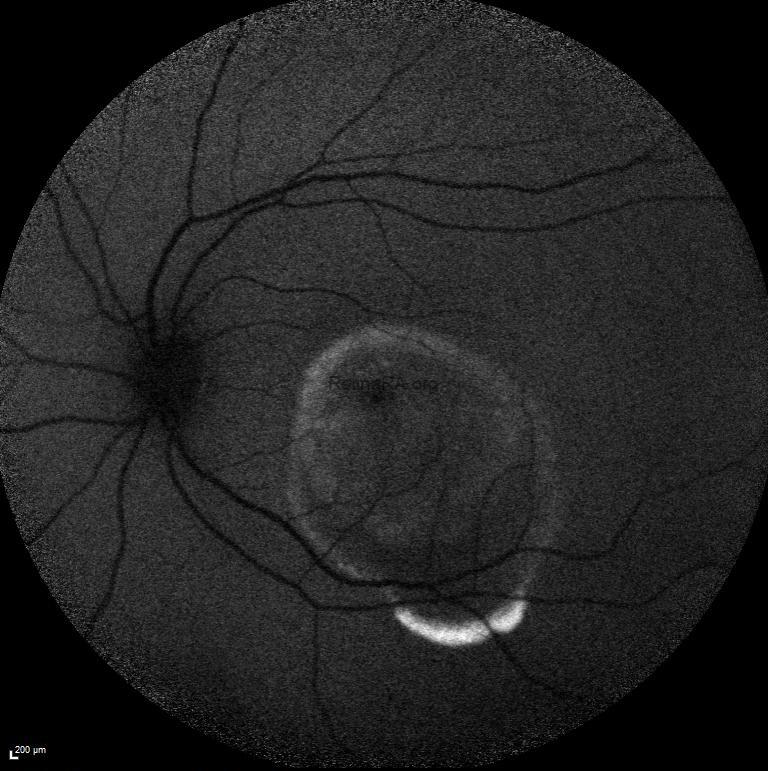
Fundus autofluoresence of both eyes showed symmetrical hypoautofluorescence with a rim of hyperauotofluorescence extending into the inferior macula of both eyes.

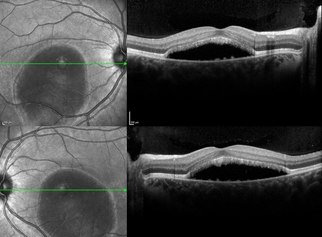
Spectral-domain optical coherence tomography illustrated hyporeflective serous macular detachment with vitelliform deposits along the floor in both eyes.
In light of these multimodal imaging findings, the preliminary diagnosis was Best disease and the diagnosis was confirmed with electrooculogram and genetic analysis.
The natural history of Best disease can progress through five possible stages, including pseudohypopyon (Stage III) and vitelliruptive (Stage IV) stages, which might be notable for serous macular detachment and large subretinal hyporeflective spaces observed under spectral-domain optical coherence tomography. The presence of serous macular detachment may lead the practitioner to assume that an exudative process is present, thus leading to unnecessary and inappropriate treatment.
The diagnosis of Best disease should be considered in patients presenting with serous macular detachment and recalcitrant subretinal fluid masquerading as neovascular age-related macular degeneration or chronic central serous chorioretinopathy to avoid unnecessary treatment procedures.
Credit: Kemal Tekin, M.D., from Ulucanlar Eye Training and Research Hospital
Instagram accounts: @retina.academy and @dr.kemaltekin