
A 15–year-old girl was presented with the decreased vision and central scotoma in her left eye for four weeks. There was no family history of eye disorders and she was not the product of a consanguineous marriage. However, it has been learnt that her mother contracted rubella during her pregnancy. Blood tests had run and serum studies showed IgM and IgG antibodies that were positive for rubella. Moreover, the maternal immunity status was not known. However, neither ocular nor systemic abnormalities were detected at birth. The patient’s physical examination revealed patent ductus arterious in the later years of her childhood. Moreover, she had a history of hearing decrease since she was one year old.
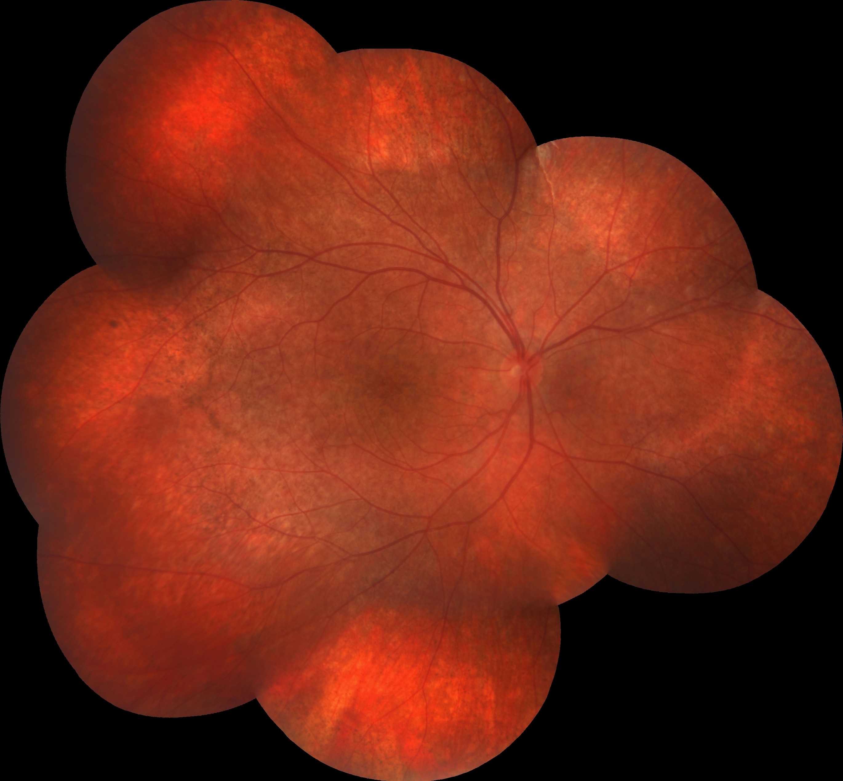
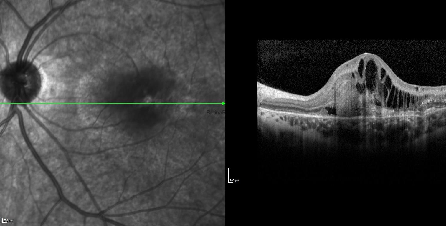
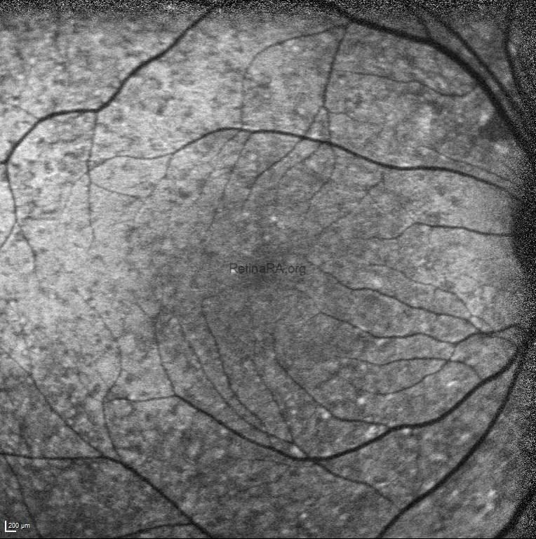
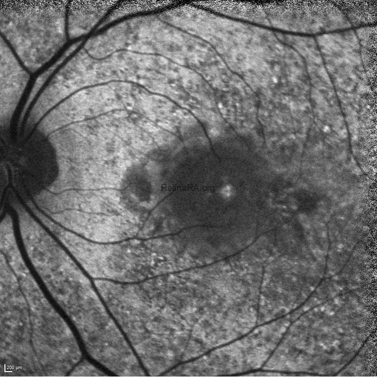
On ocular examination, the BCVA was 20/20 in the right eye and 20/400 in the left eye. IOPs were within normal limits in both eyes and anterior segment examinations were unremarkable. Dilated fundus examination of the right eye revealed a normal optic disk with a classical rubella retinopathy findings — a classic salt-and-pepper appearance of the retina that is due to the distribution of areas of increased and decreased pigmentation. In the left eye, in addition to classical rubella retinopathy findings appearance, a whitish subretinal lesion with retinal hemorrhages in the macula was detected. Mottling of the RPE was also evident in both eyes.

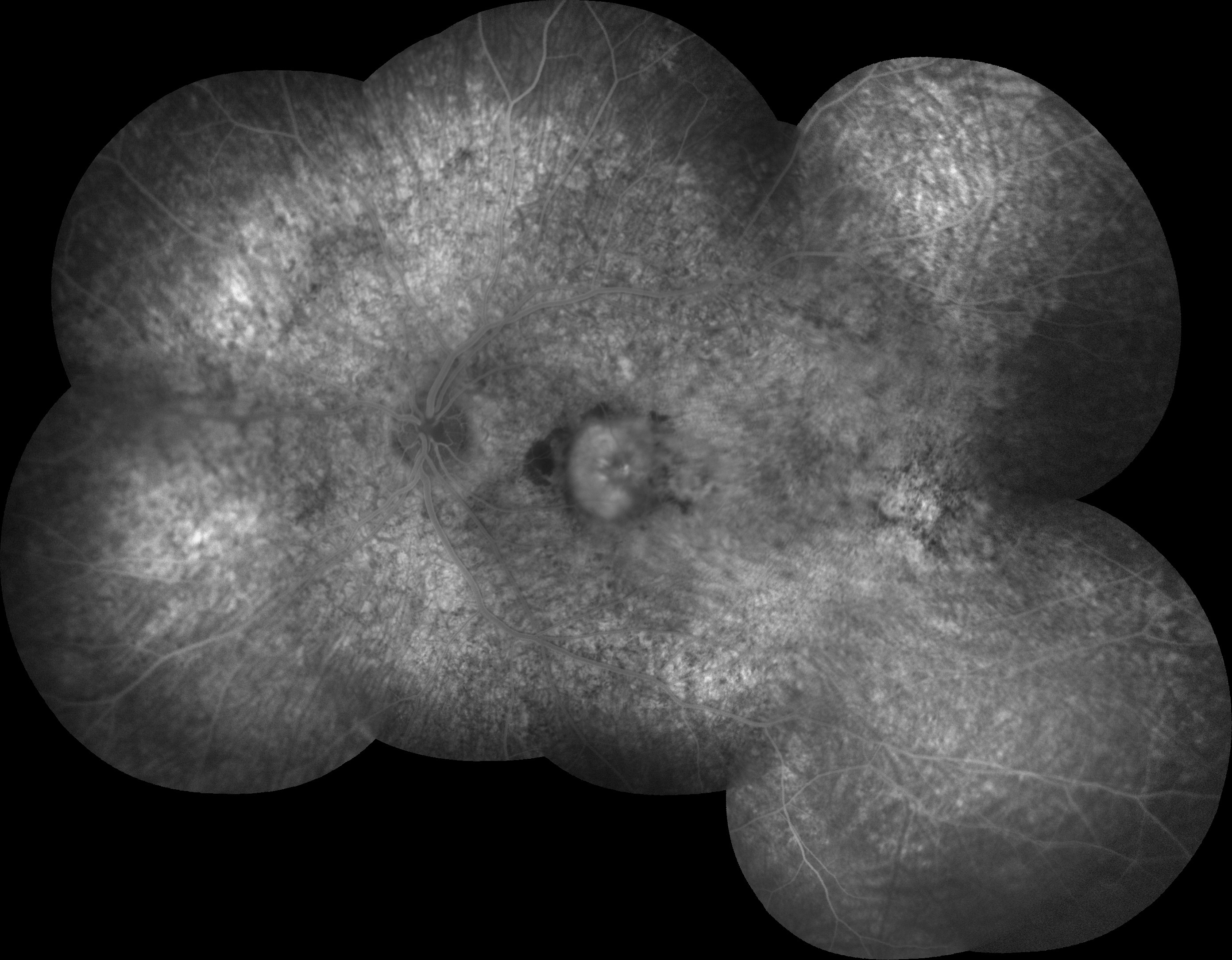
Fundus fluorescein angiography illustrated a pattern of diffuse spotty fluorescence because of the defective RPE without any leakage or staining in the right eye. On the left eye, in addition to diffuse spotty fluorescence, an active subfoveal CNV lesion, that hyperfluoresces in the early phases of the FFA, maintains well-demarcated borders, and leaks, was detected.
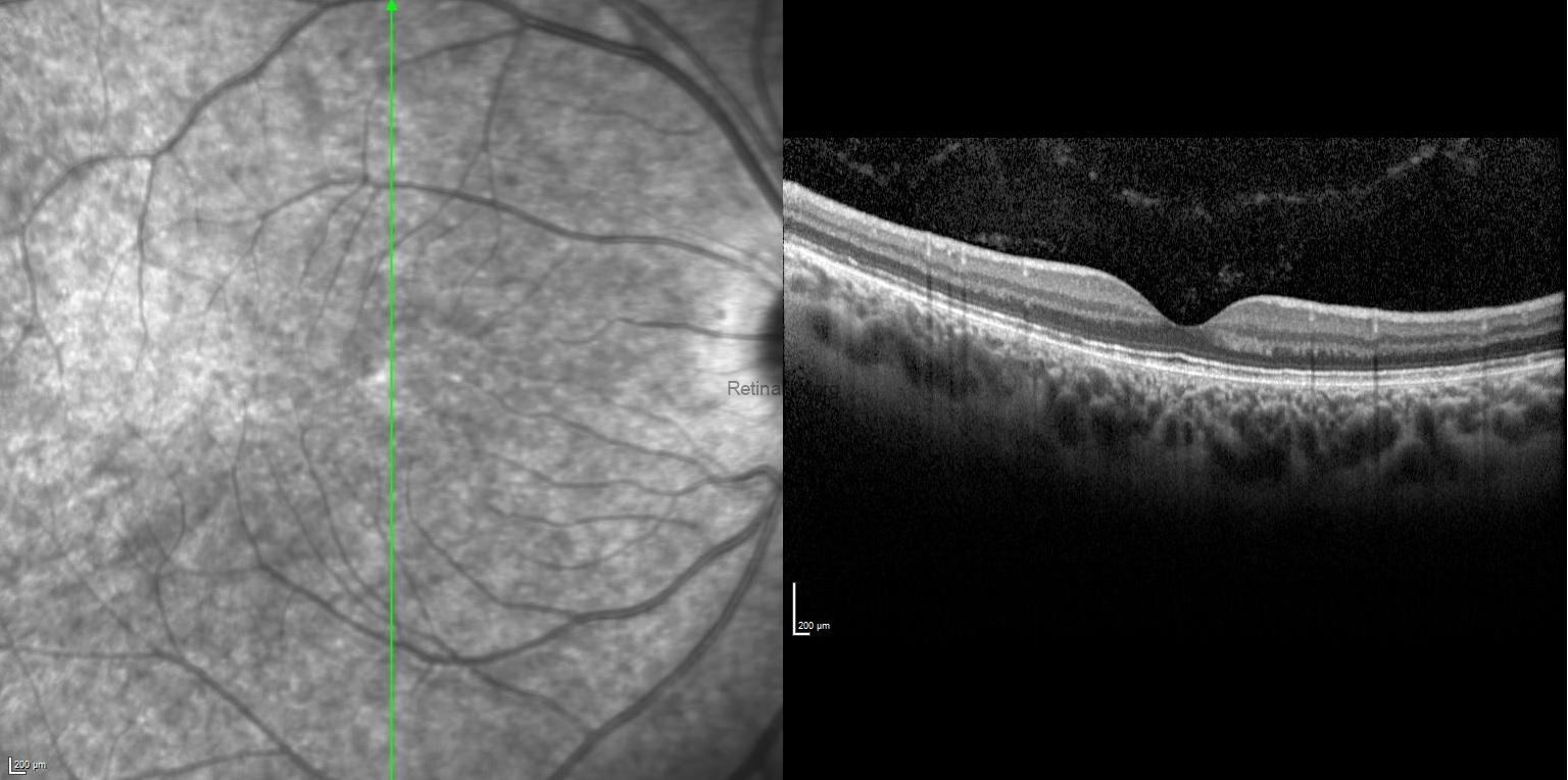
SD-OCT of the right eye showed attenuated inner segment–outer segment junction, absent upward displacement of subfoveal ellipsoid zone band, and mottled retinal pigment epithelium, while it revealed thickened and elevated retinal layers at the macula due to the subretinal and intraretinal fluid with foveal and extrafoveal protruding hyper-reflective material in the left eye.
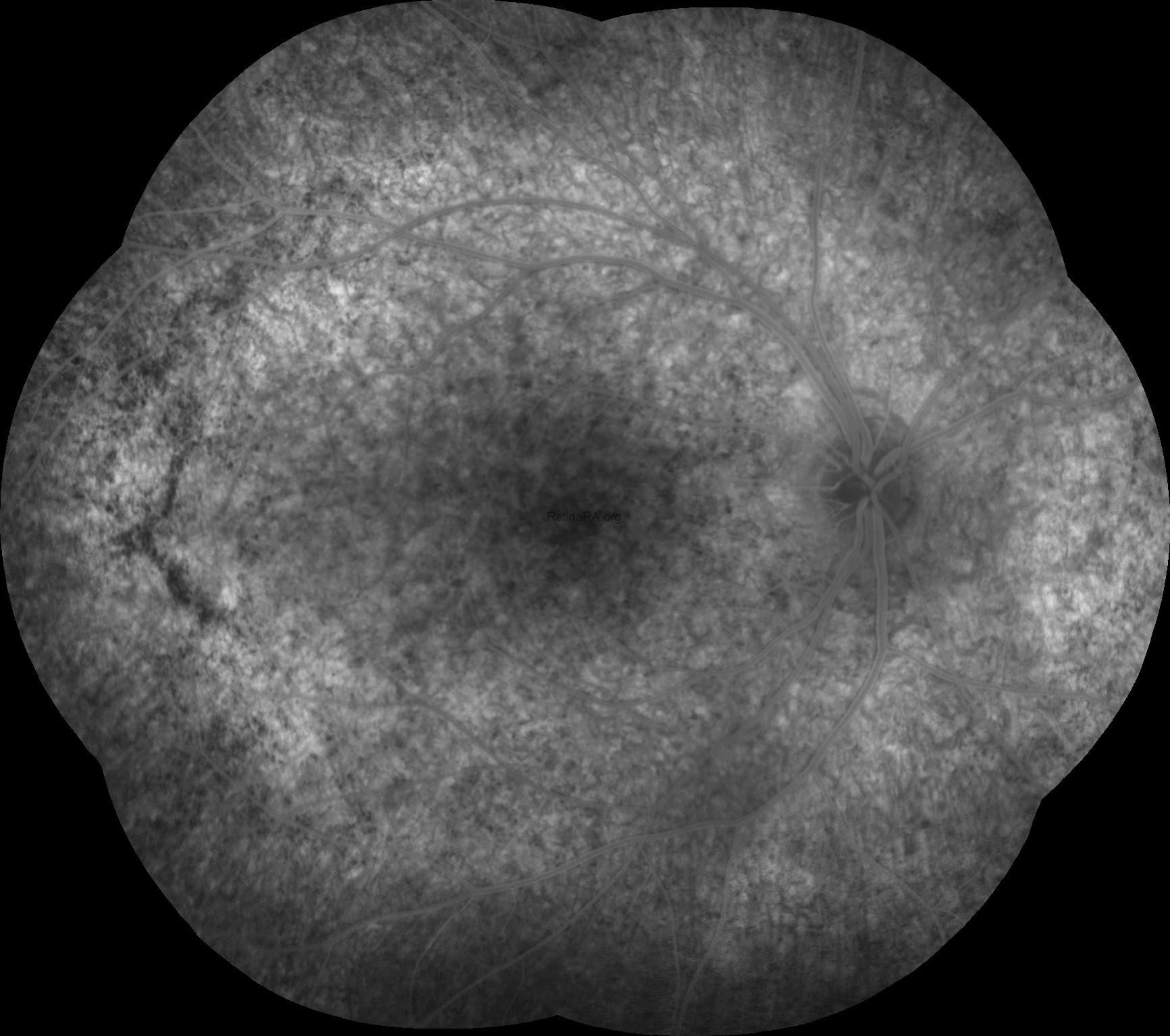
Fundus autofluorescence highlighted the fundus abnormalities patchy auto-fluorescence with a stippled hypo-fluorescence in both eyes, and also an hypo-autoflorescence area corresponding to the area of choroidal neovascularization in the left eye.
Credit: Kemal Tekin, M.D., from Ulucanlar Eye Training and Research Hospital
Instagram accounts: @retina.academy and @dr.kemaltekin