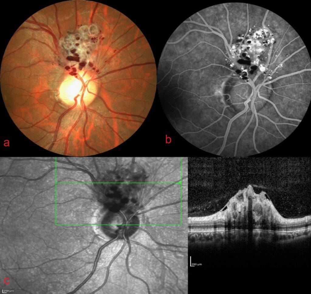
A 49-year-old female was referred to the retina clinic because of a peripapillary vascular tumor which was detected during routine eye examination. Fundus examination showed peripapillary grape-like clusters of red and dilated sac-like aneurysms filled with dark red blood.

Fundus fluorescein angiography revealed typical fluorescein-blood levels indicating the plasma-erythrocytic separation within some aneurysms due to pooling of dye in the superior plasma (hyperfluorescent) and sedimented red blood cells inferiorly (hypofluorescent).

Spectral-domain optical coherence tomography passing through the lesion exhibited the presence of grape bunch shaped multiple hyporeflective vesicular formations surrounded by a hyperreflective edged-ring involving inner retinal layers.

Retinal cavernous hemangioma is seen along the course of major veins or in the peripapillary area with aneurysmal dilation of venules. It might be associated with cutenous or central nervous system hemangiomas. The diagnosis can be made based on the distinctive appearance of the fundus, combined with imaging findings.
Credit: Kemal Tekin, M.D., from Ulucanlar Eye Training and Research Hospital
Instagram accounts: @retina.academy and @dr.kemaltekin
