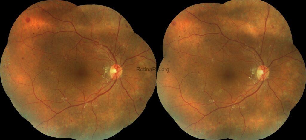
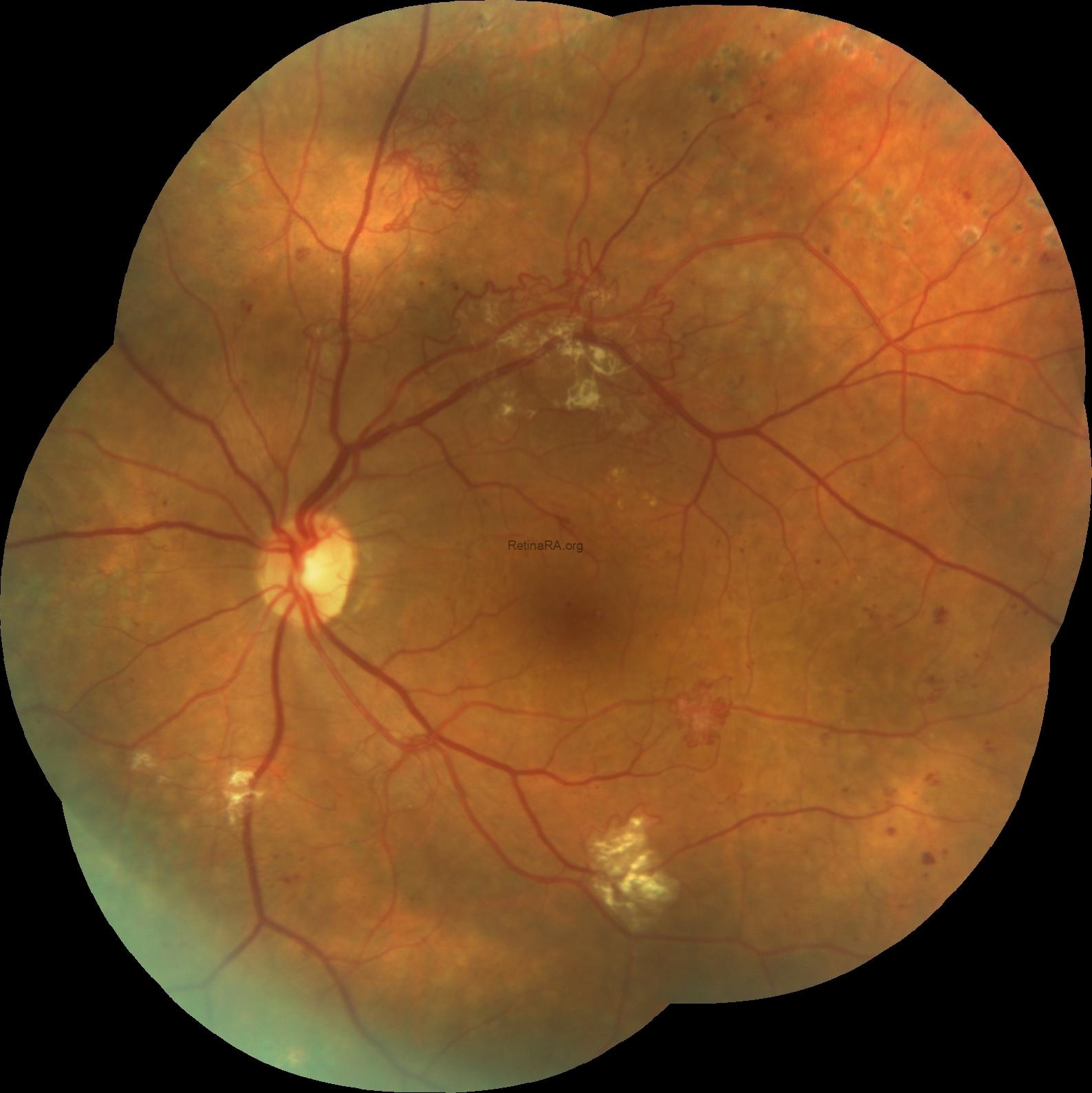
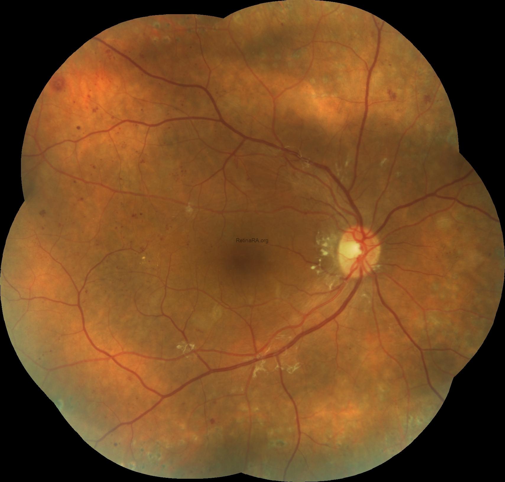
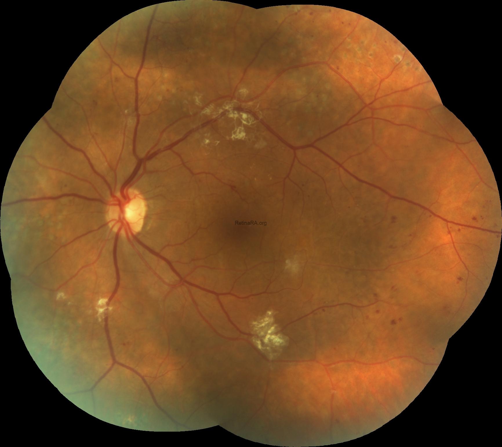
This was a 40 year old female who have poor-controlled type 1 diabetes. The BCVAs were 20/20 for both eyes and anterior segment examinations were unremarkable.
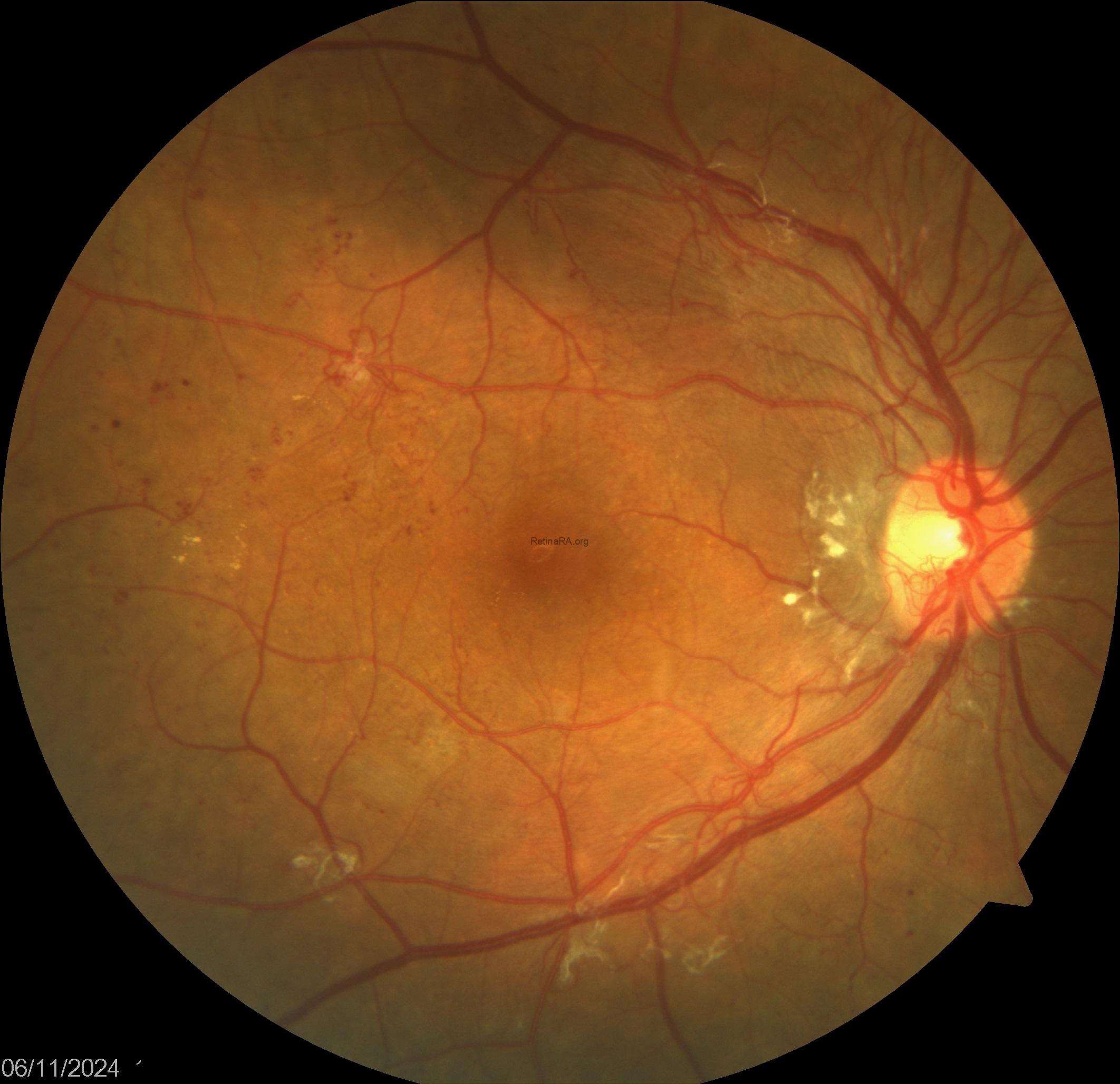
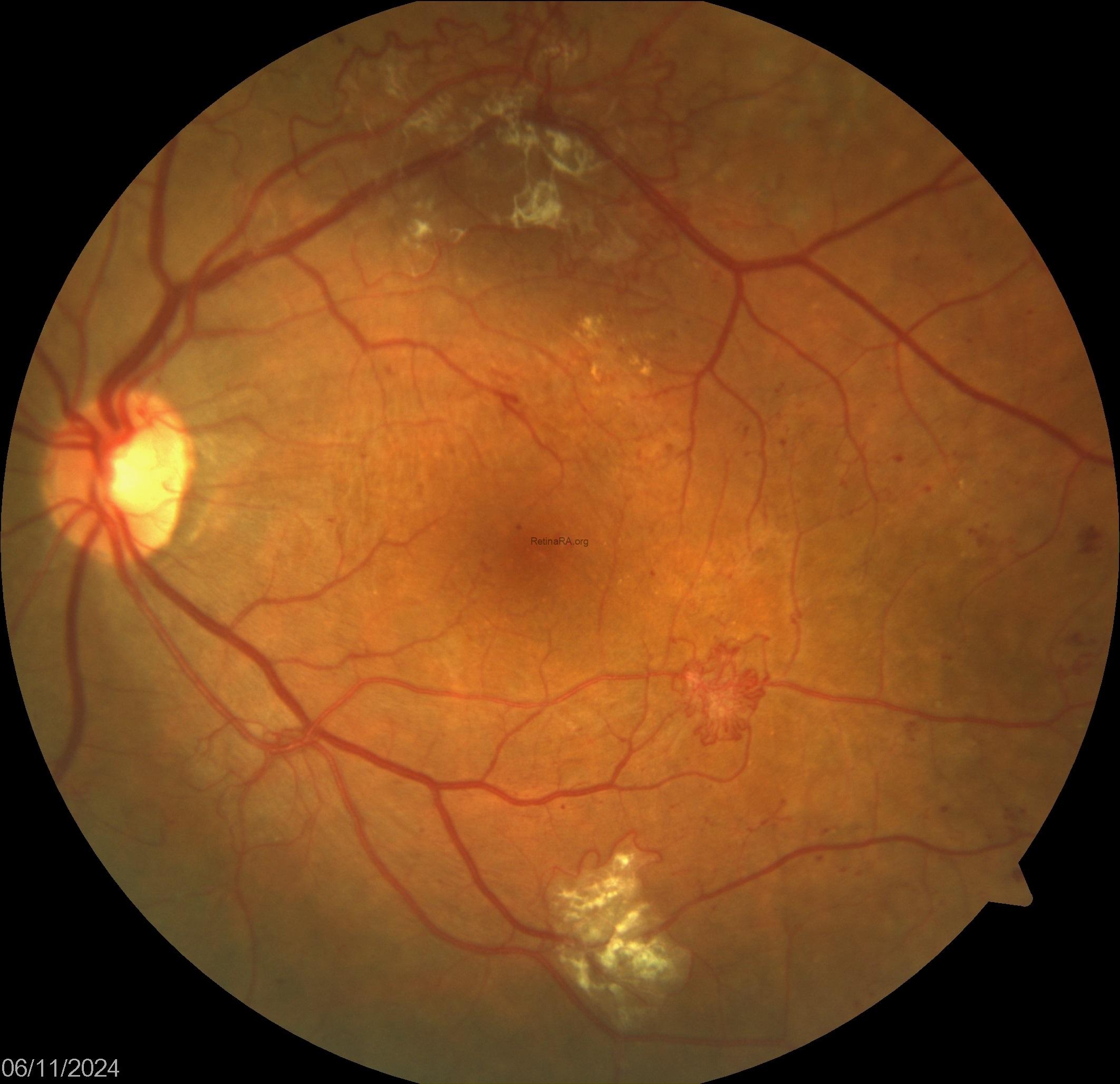
Fundus images showed widespread retinal neovascularization in both eyes, optic nerve neovascularization in the right eye and fibrovascular proliferation in the left eye. The previous laser photocoagulation spots were olso seen in the midperipheral retina of both eyes. However, it was noticed that peripheral retinal areas were empthy in both eyes without any laser spots.
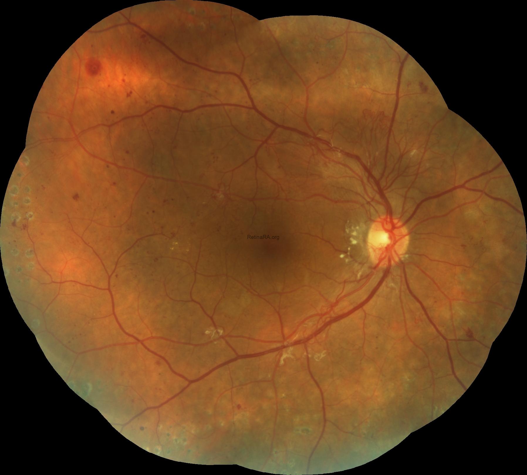
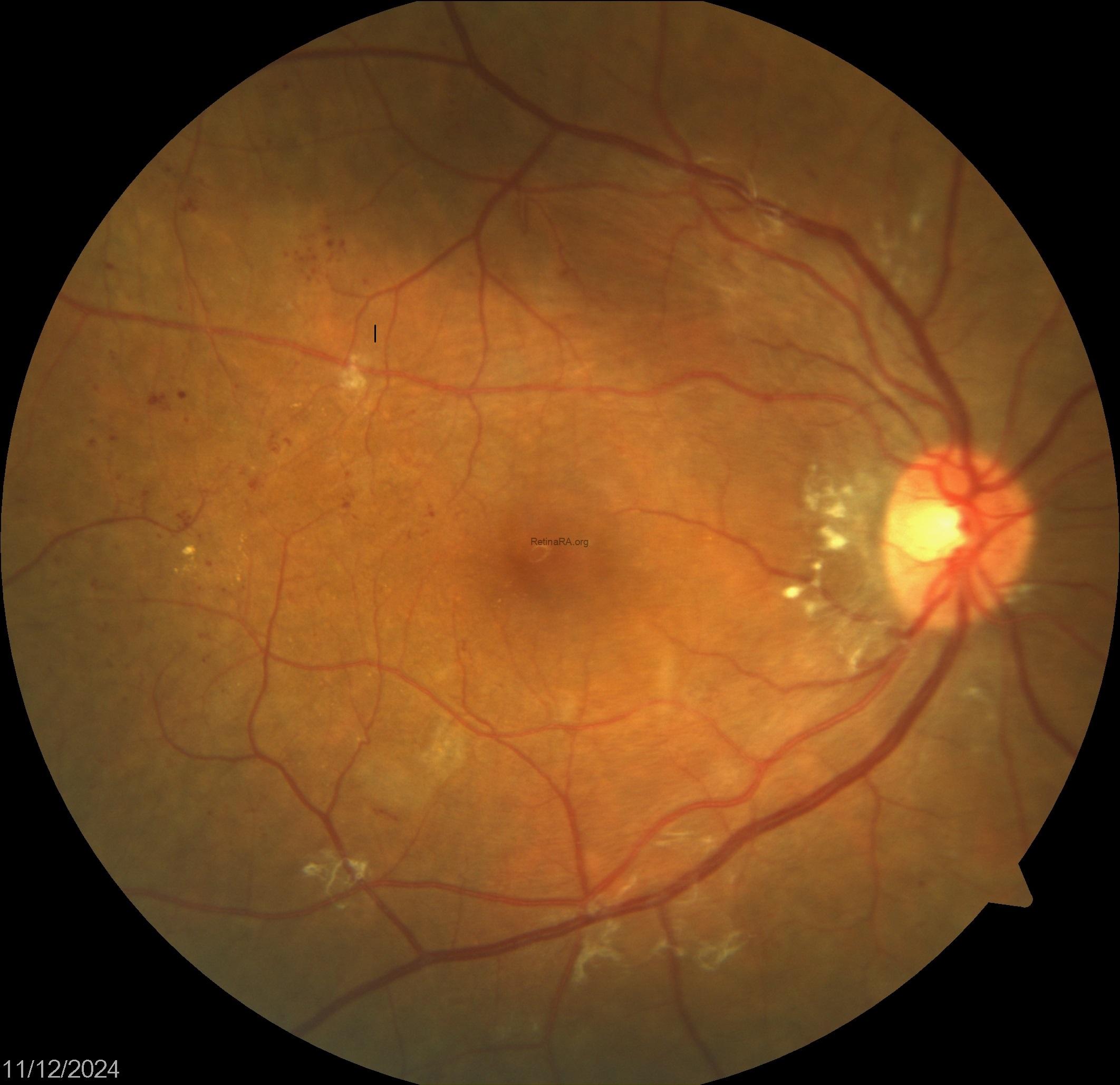
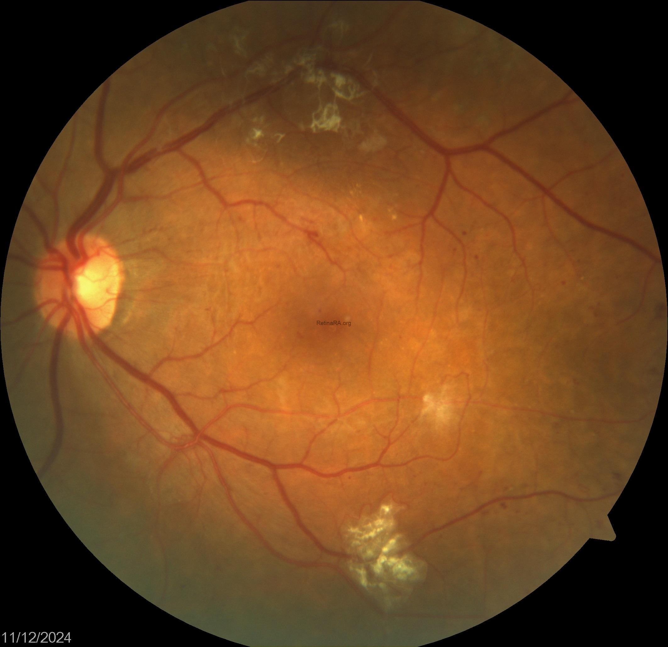
Peripheral retinal panretinal laser photocoagulation were applied and just after completing laser treatment, a single dose of anti-VEGF injection were administered to both eyes of the patient. At the first-month follow-up visit after the treatment, it was observed that all neovascularization had completely regressed.
Proliferative diabetic retinopathy (PDR) represents a significant therapeutic problem that often leads to severe visual loss. Panretinal photocoagulation (PRP) has long been a mainstay treatment for PDR. Conversely, intravitreal anti-VEGF therapy has served as an alternative treatment for PDR. It was showed that PRP combined with anti-VEGF therapy is superior to PRP alone in the management of PDR. This combination treatment yields better and faster regression of neovascularizations and a lower incidence of serious complications requiring pars plana vitrectomy. To achieve regression of neovessels, anti-VEGF therapy has a potential role in select cases of laser-treated PDR cases without any evidence of traction.
Credit: Kemal Tekin, M.D., from Ulucanlar Eye Training and Research Hospital
Instagram accounts: @retina.academy and @dr.kemaltekin