
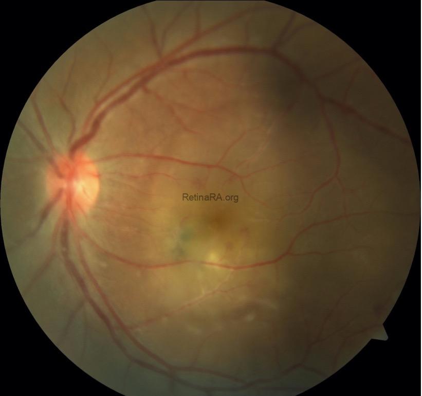
A 23y old, female patient presented with decreased vision in her left eye. Best corrected visual acuity levels were 1.0/CF 5, intraocular pressures were within normal limits, and there was 1+ anterior chamber reaction in her left eye. On fundus examination, while the right eye was normal, a hyperpigmented scar and active lesion were observed near the fovea in the left eye. There was involvement of the arteries adjacent to the lesion.
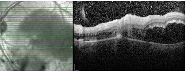
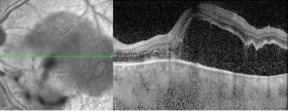
OCT showed intravitreal hyperreflective spots concentrated on the lesion, full-thickness retinal involvement, and serous retinal detachment.
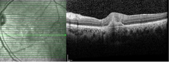
With the diagnosis of ocular toxoplasmosis, trimethoprim sulfamethoxazole with azithromycin and oral steroid treatments were started. In the 1st month control OCT imaging, scarring in the lesion and complete recovery of serous detachment were observed.
Ocular toxoplasmosis should also be considered in the differential diagnosis of serous retinal detachment. Serous retinal detachment accompanying the lesion is observed in 20-25% of toxoplasma cases, and this rate may increase even more with routine OCT imaging. Its distinguishing features are unilateral nature, typical retinochoroiditis focus and/or the presence of old scar.
Credit: Merve İnanç Tekin, MD, FEBO
Ulucanlar Eye Training and Research Hospital, Ankara, Turkey
Instagram accounts: @uveacademy and @merveinanctekin