A 30 year-old male patient admitted to clinic with a complaint of decreased vision after blunt trauma in his left eye. He felt this complaint since being punched 2 days previously. The best corrected visual acuity was 20/200 in the affected eye with a normal intraocular pressure. Anterior segment examination was unremarkable.
Dilated fundus examination of the left eye showed commotio retina, widespread intraretinal hemorrhage and fluid with macular involvement and choroidal rupture.

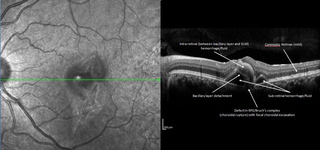
Fundus autofluorescence imaging demonstrated the hypoautofluorescence areas corresponding to intraretinal hemorrhages and hyperautofluorescence areas representing the choroidal rupture.

The spectral domain optical coherence tomography (OCT) imaging revealed defective retina pigment epithelium-Bruch’s membrane complex with a focal choroidal excavation, mild commotio retina, cystic subretinal hemorrhage areas, bacillary layer detachment and intraretinal hemorrhages that located between bacillary layer and external limiting membrane.
OCT angiography also showed the defective retina pigment epithelium-Bruch’s membrane complex confirming the choroidal rupture without choroidal neovascularization.

OCT made possible noninvasive imaging of the microscopic anatomy of the retina, a nearly transparent structure. With the improvement in OCT imaging technology, photoreceptor inner segments are divided into two parts, the myoid and the ellipsoid zones. The myoid has ribosomes, some endoplasmic reticulum, golgi bodies, and rare mitochondria while the ellipsoid is densely packed with mitochondria.
Bacillary layer detachment (BALAD) is an OCT finding that has been reported in many uveitic and retinal diseases. It describes the splitting at the level of the inner segment myoid of photoreceptors leading to development of intraretinal fluid cavities. There is a separation of the bacillary layer from the other retinal layers because of a split at the photoreceptors immediately posterior to the external limiting membrane within the photoreceptor inner segment myoid. The intraretinal fluid is bounded by the myoid and externally by the ellipsoid zone and photoreceptor outer segments, the latter two usually remain attached to the retinal pigment epithelium.
Credit: Kemal Tekin, M.D., from Ulucanlar Eye Training and Research Hospital
Instagram accounts: @retina.academy and @dr.kemaltekin
