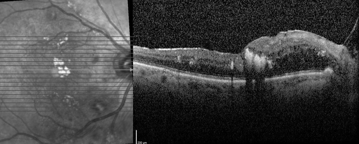
A 65-year-old female diagnosed with type 2 diabetes mellitus and impaired lipid metabolism with hypercholesterolemia presented with moderately graded non-proliferative diabetic retinopathy with widespread hard exudates (HEs) at the macula (Figure 1, top). The HEs appeared as golden-yellow lesions at the fovea and pale yellow-colored HEs surrounding the foveal lesion were also detected in temporal and superior to the fovea (Figure 1, top). Spectral domain optical coherence tomography (SD-OCT) scans through the fovea showed serous macular detachment and nasally cystoid macular edema in addition to an unusual finding of intensely hyper-reflective line deposits over the retina pigment epithelium without posterior shadowing which was termed as Onion Ring Sign. (Figure 1, middle-bottom). Two months after injection of dexamethasone implant (Ozurdex®; Allergan, Inc., Irvine, CA, USA; 700 μg) in addition to systemic lipid lowering therapy, cystoid macular edema and serous retinal detachment disappeared besides the onion ring sign resolved as subfoveal exudate plaque (Figure 2). However, there was not any increment in visual acuity.
In the eyes having macular neovascularization, the cholesterol crystals are seen as horizontal hyper-reflective deposits in the subretina pigment epithelium-basal laminar space, and appear as highly intense signals without any shadowing on OCT. It was revealed that horizontal hyper-reflective deposits on OCT in diabetic retinopathy which were different from the HEs showing posterior shadowing. These were described as hyper-reflective crystalline deposits and were termed as the “onion ring sign.
Credit: Kemal Tekin, M.D., from Ulucanlar Eye Training and Research Hospital
Instagram accounts: @retina.academy and @dr.kemaltekin
Composite fundus photo and OCT scans of the patient with type 2 diabetes mellitus and hypercholesterolemia.
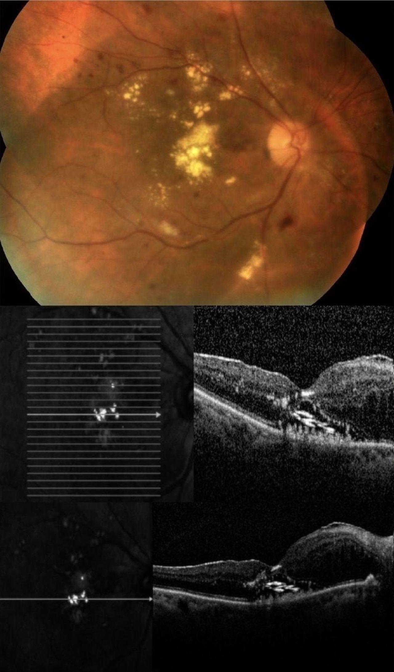
OCT scan of the patient two months after injection of dexamethasone implant.