A 48-year-old male patient presented to the outpatient clinic for a routine eye examination. Best-corrected visual acuity was 20/20 in both eyes, and intraocular pressures were within normal limits. Anterior segment examination was unremarkable.
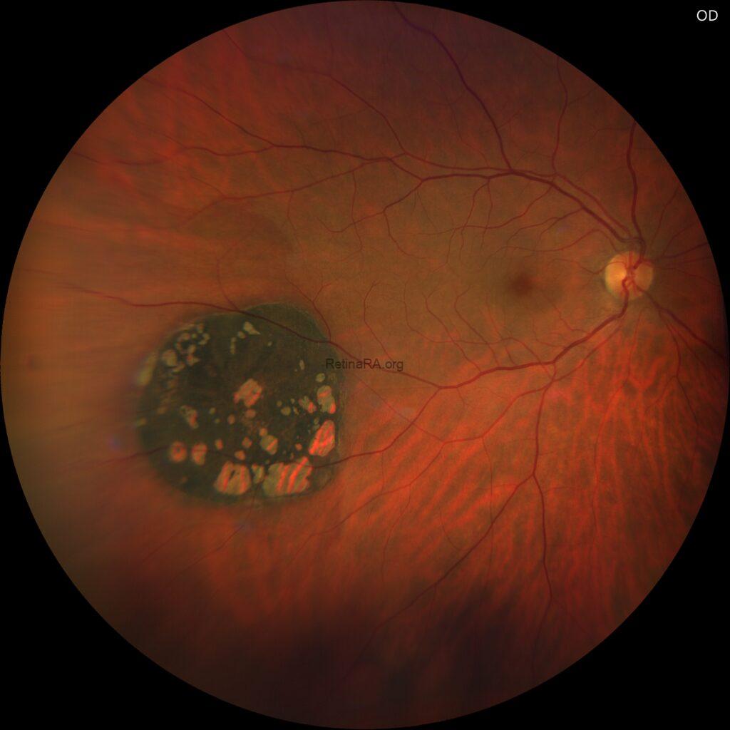
Fundus examination of the right eye revealed a solitary, flat, round, darkly hyperpigmented retinal lesion with multiple hypopigmented lacunae in the mid-peripheral inferotemporal quadrant, whereas the left eye appeared completely normal.


Solitary congenital hypertrophy of the retinal pigment epithelium (CHRPE) is a benign, congenital retinal lesion often found incidentally during routine eye examinations. It usually presents as a solitary, flat, round or oval, well-defined darkly pigmented lesion, most often located in the mid-peripheral fundus of one eye. Characteristic features may include small hypopigmented lacunae or scalloped edges within the lesion. Importantly, solitary CHRPE remains stable over time, is asymptomatic, and does not impair visual function. Unlike multiple CHRPE lesions, which can occasionally be linked to systemic conditions such as Gardner syndrome, the solitary form has no systemic associations. Management is conservative, requiring only documentation and periodic monitoring without any active treatment.
Credit: Kemal Tekin, M.D., from Ulucanlar Eye Training and Research Hospital
Instagram accounts: @retina.academy and @dr.kemaltekin
