A 30-year-old male presented with progressive vision loss in his left eye. His history was notable for prior resection of a cervicomedullary hemangioblastoma, with no ophthalmic follow-up afterward.
BCVA was 1.0 OD and counting fingers OS. Fundus examination of the right eye showed a small peripheral retinal capillary hemangioblastoma (1.2 DD) with mildly dilated feeder and draining vessels. The left eye demonstrated a large 6 DD hemangioblastoma at the equator, accompanied by extensive subretinal and intraretinal exudation, macula-involving exudative retinal detachment, and markedly tortuous feeder vessels. Additional systemic imaging revealed pancreatic and renal cysts, supporting the diagnosis of Von Hippel–Lindau disease.
Laser photocoagulation achieved long-term stability in the right eye. The left eye underwent periocular corticosteroid therapy, cryotherapy, and subsequently pars plana vitrectomy with tumor excision and silicone oil tamponade, later followed by cataract extraction. Final VA remained 0.02. (The authors reported that postoperative images of the left eye were not available.)
Credit: Akhmetova Zhanara, MD. Rassuliyeva Meruyert, MD.
Kazakh Research Institute of Eye Diseases. Almaty. Kazakhstan.
Instagram account: @ah_janara, @rasulieva
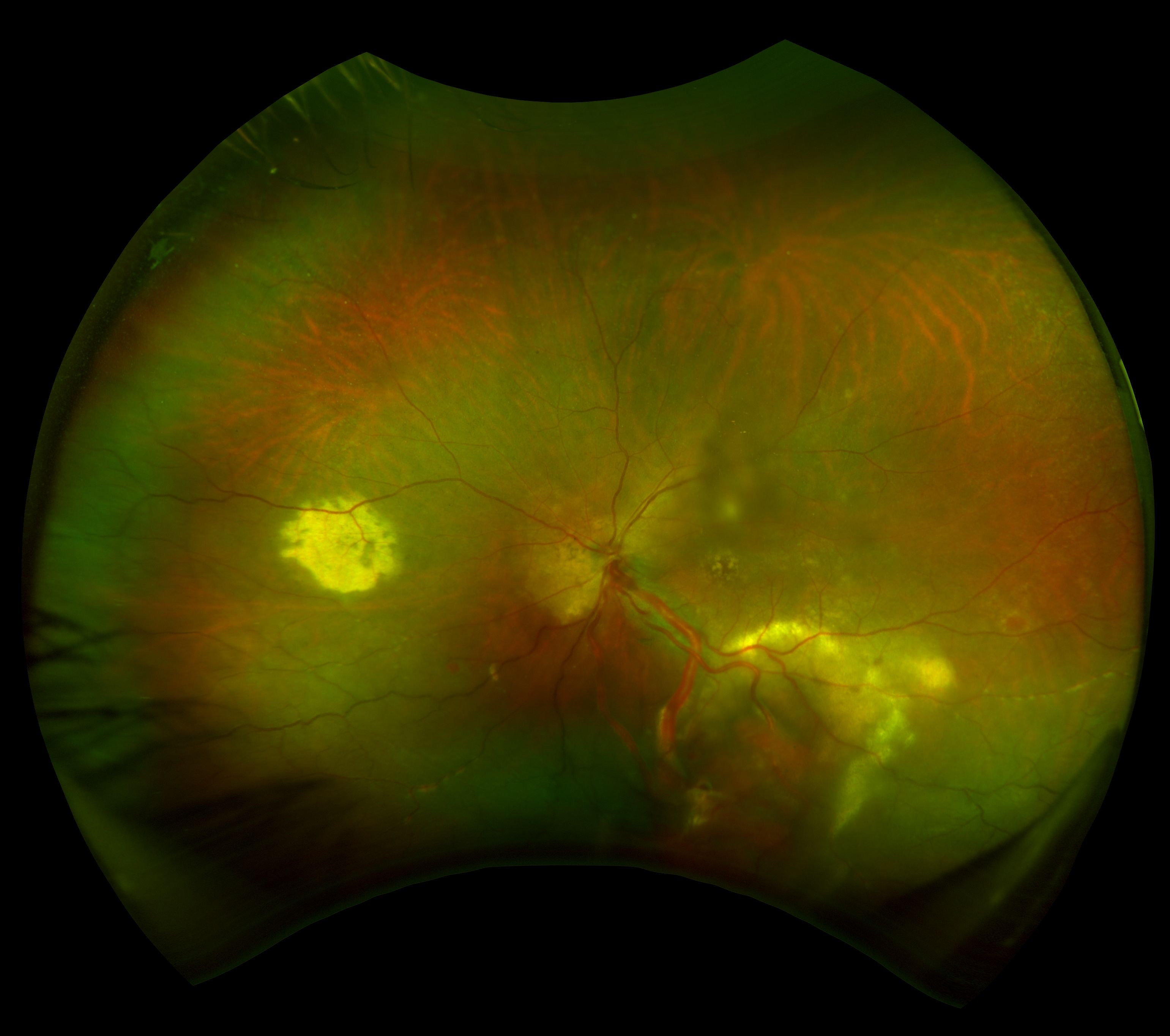
The wide-field image of the right eye shows a small peripheral retinal capillary hemangioblastoma.
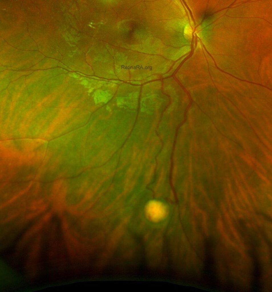
Post-laser photocoagulation image of the right eye with a small peripheral retinal capillary hemangioblastoma.
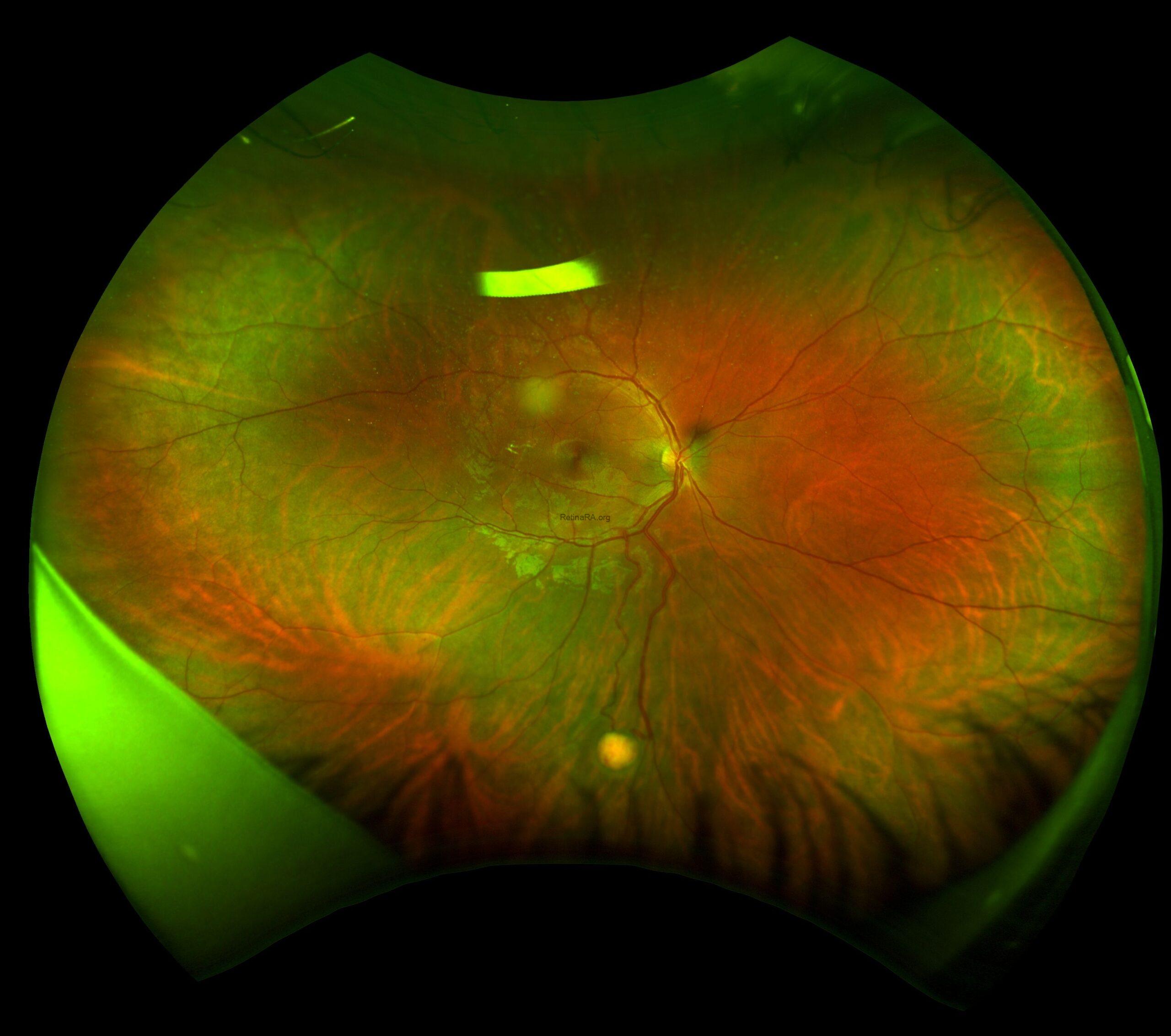
 Preoperative image of the left eye with a large 6 DD retinal capillary hemangioblastoma.
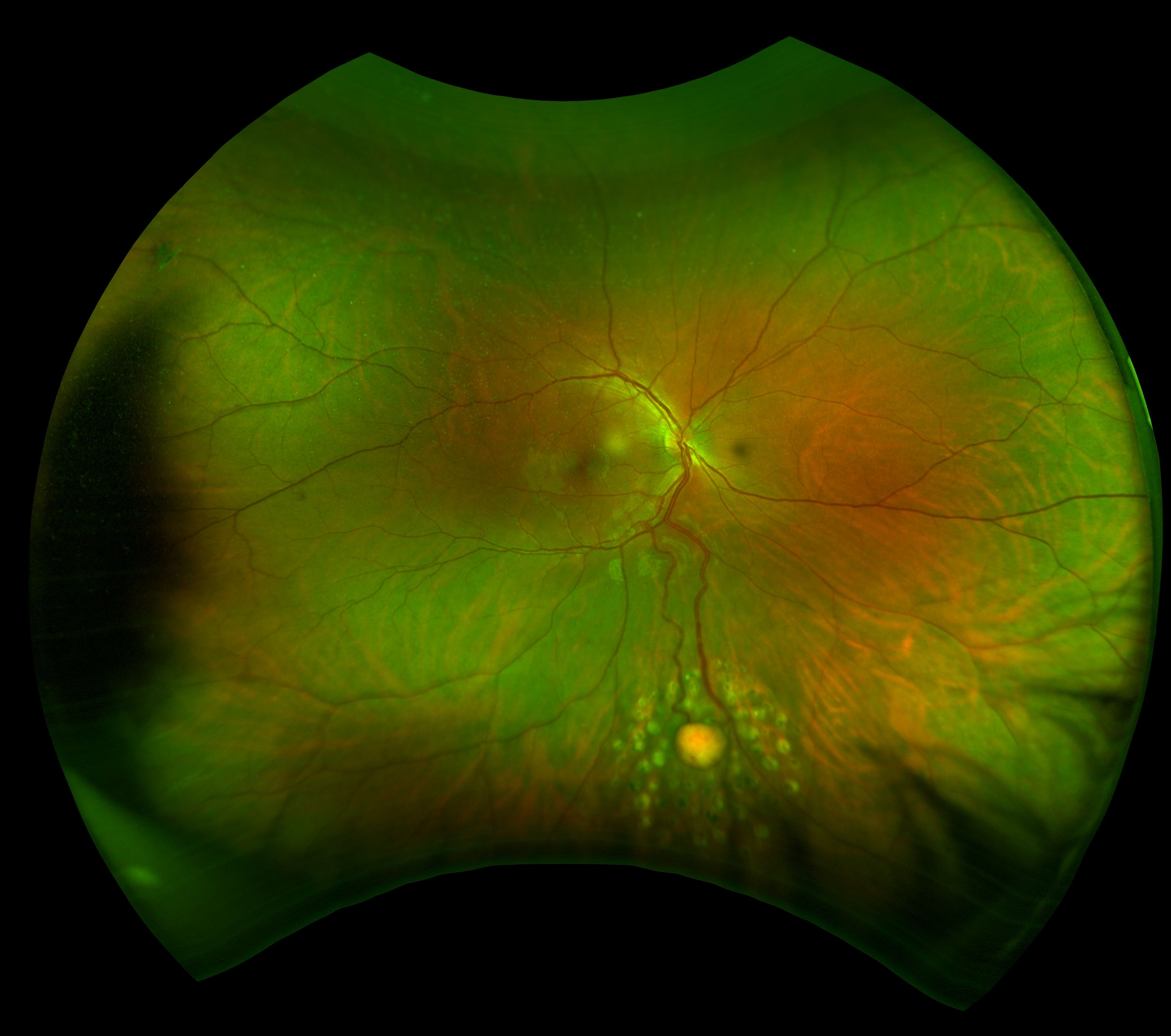