A 27-year-old male patient applied for routine eye examination. He underwent three intravitreal injections in the right eye 2 year ago and had no history of previous ocular surgery. On ocular examination, the BCVAs were 20/200 with the correction of -12.00 diopters in the right eye and 20/30 with the correction of -14.00 diopters in the left eye. Intraocular pressures were within normal limits in both eyes and no pathology was observed in the anterior segment examination.
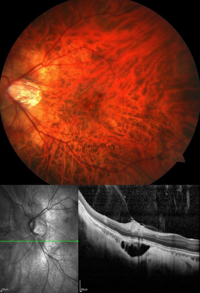
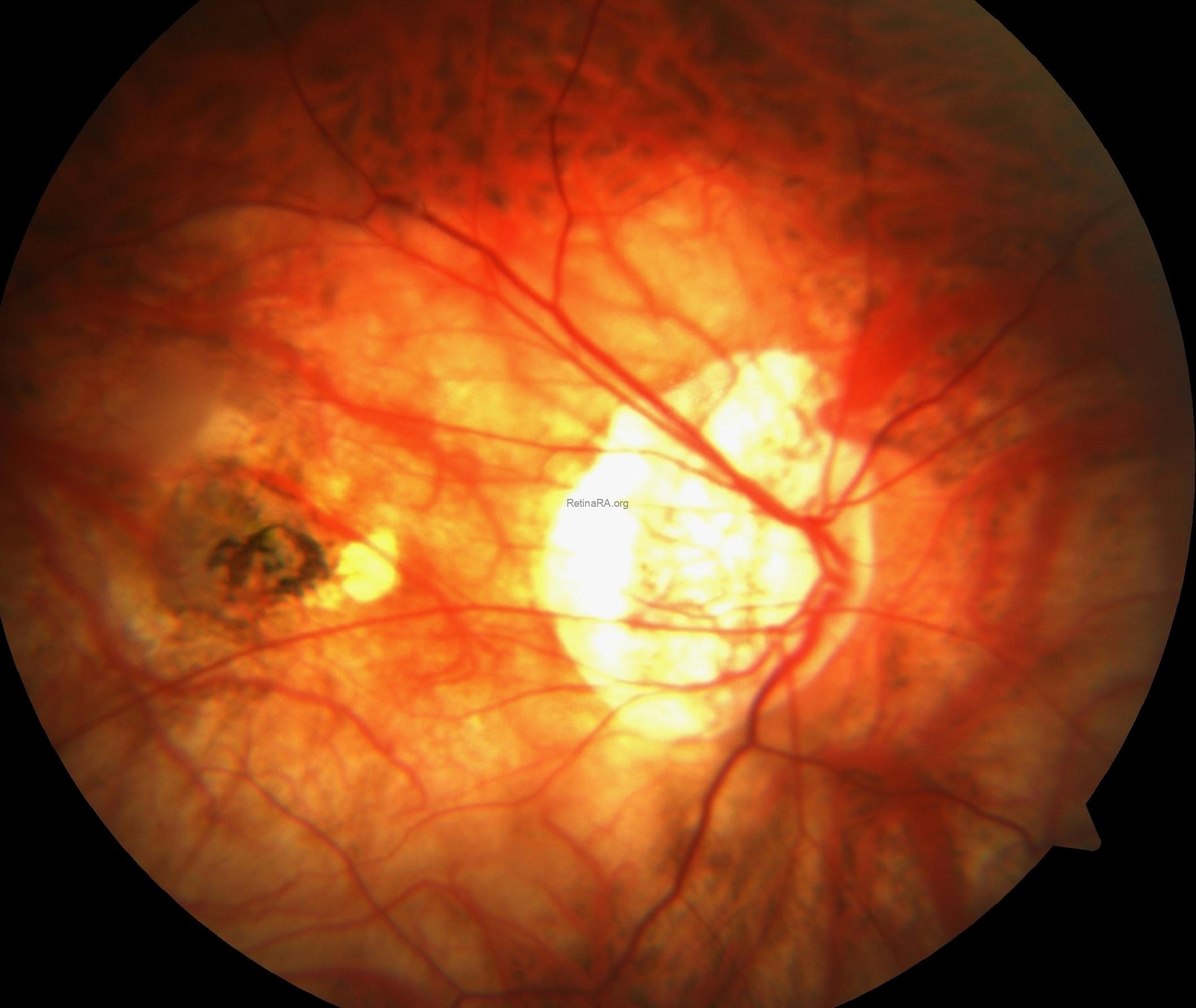
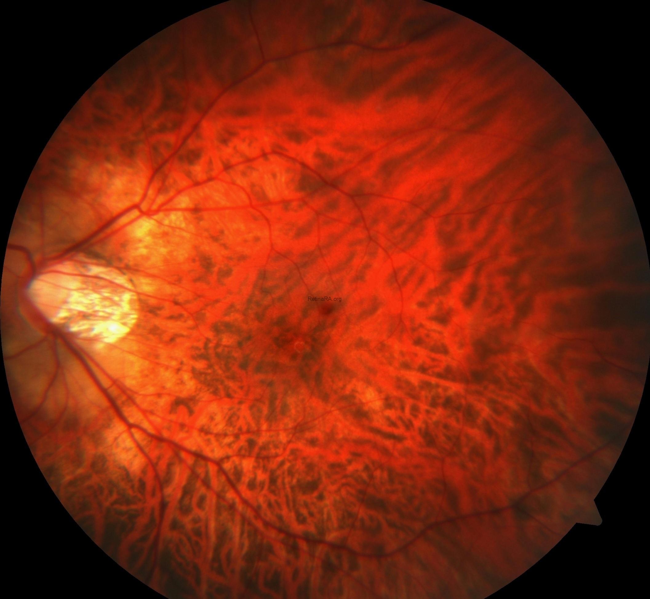
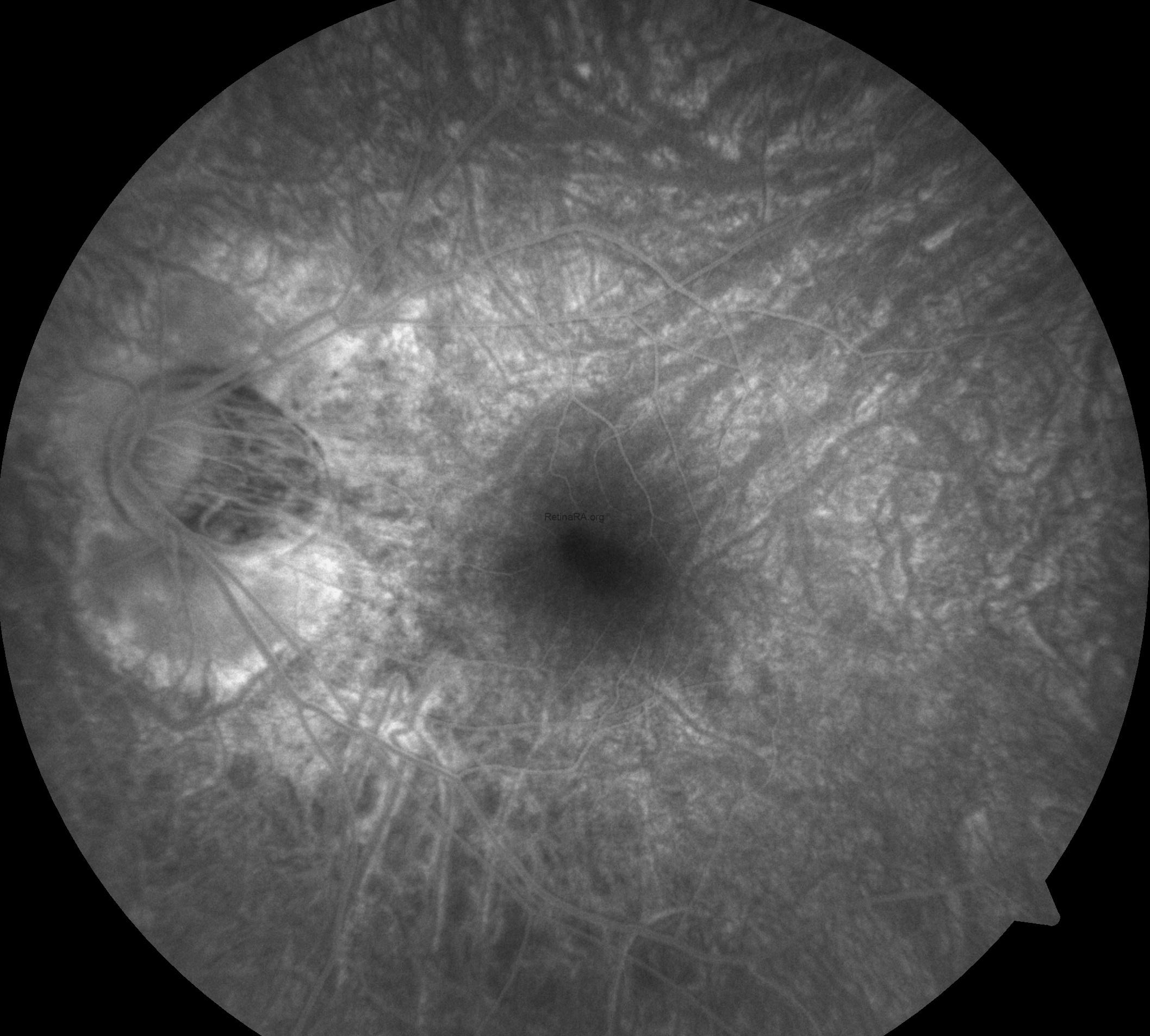
Dilated fundus examination of the right eye showed tilted disc, peripapillary and chorioretinal atrophy as well as macular scar, while the left eye exhibited aa well-defined, yellow-orange lesion located inferiorly along the border of the myopic crescent in additon to tilted disc, peripapillary and chorioretinal atrophy.
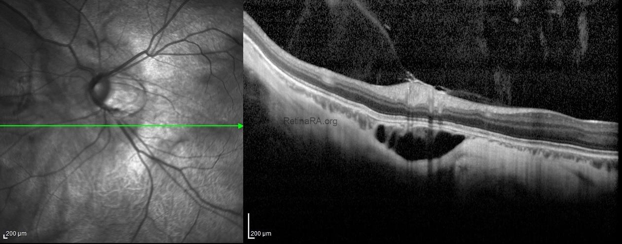
Spectral-domain optical coherence tomography showed the choroidal neovascular membrane scar without any subretinal or intraretinal fluid in additon to posterior staphyloma in the right eye. For the left eye, the OCT-scan passing throught the lesion under the optic disc exhibited an intrachoroidal space with a hyporeflective appearance below the normal plane of retinal pigment epithelium with normal Bruch’s membrane and retinal layers over the lesion.

Fundus fluorescence angiography showed scar staining without leakage in the right eye, while the peripapillary lesion in the left eye appeared as hypofluorescent in the early stages of angiography and hyperfluorescent without leakage in the late stages.

Peripapillary intrachoroidal cavitation is a yellow-orange lesion which located at the outer border of the myopic conus. First described as a localized detachment of the retinal pigment epithelium, its intrachoroidal location was later revealed, justifying its current name. It can be seen with other myopic complications such as posterior staphyloma, but its pathogenesis is not still clear to. Although it has been considered a benign condition, most eyes with intrachoroidal cavitaiton demonstrate visual field defects leading to diagnostic uncertainty as these deficits resemble those seen in glaucomatous eyes. With the advances in optical coherence tomography, high optic nerve sheath traction forces during eye movements in highly myopic eyes have been suggested as promoters of intrachoroidal cavitaiton.
Credit: Kemal Tekin, M.D., from Ulucanlar Eye Training and Research Hospital
Instagram accounts: @retina.academy and @dr.kemaltekin