A 34-year-old male patient was applied to ophthalmology clinic due to transient visual obscurations. It was also learnt that he was suffering from headaches and pulsatile tinnitus for approxiately 1-month. He has no known systemic diseases and was not on any systemic or ocular drugs. The BCVAs were 20/20 for both eyes and IOPs were within normal limits. Anterior segment examination was unremerkable.
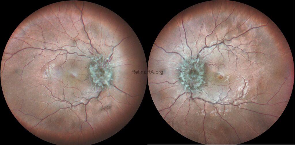
Multicolor and infrared fundus images revealed bilateral severe optic disc swelling, elevation of the disc with blurred margins, smoothly demarcated peripapillary halo, optic nerve head hyperemia, peripapillary hemorrhages, and obscuration of cup and major vessel (Grade 5 according to Frisén Scale).
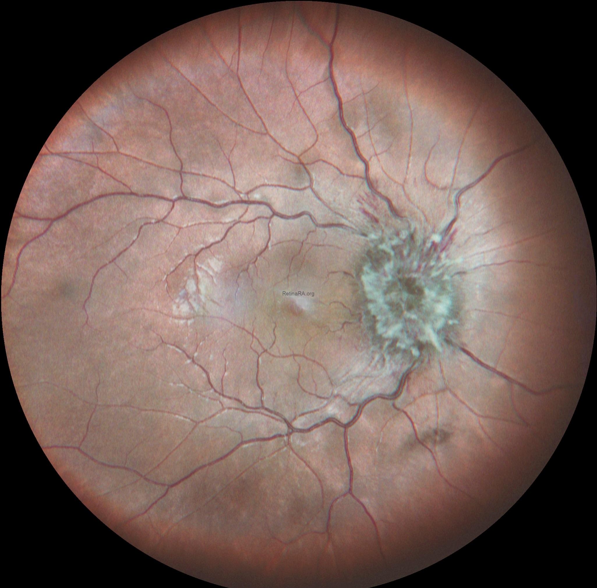
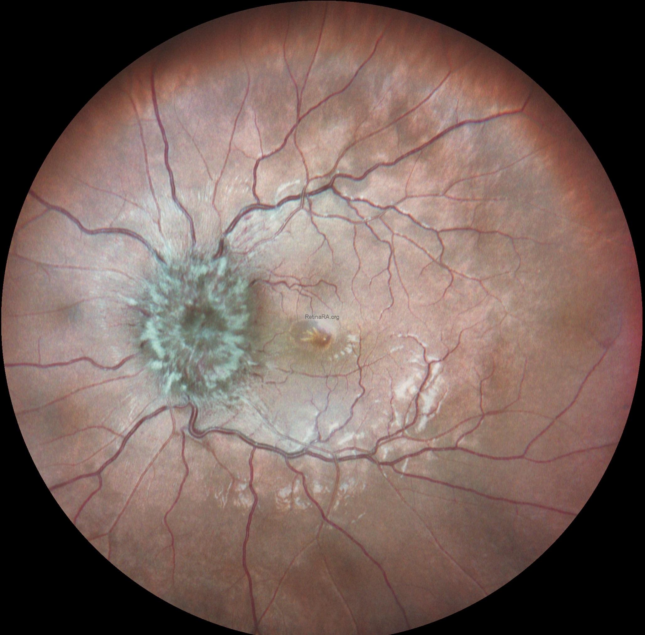
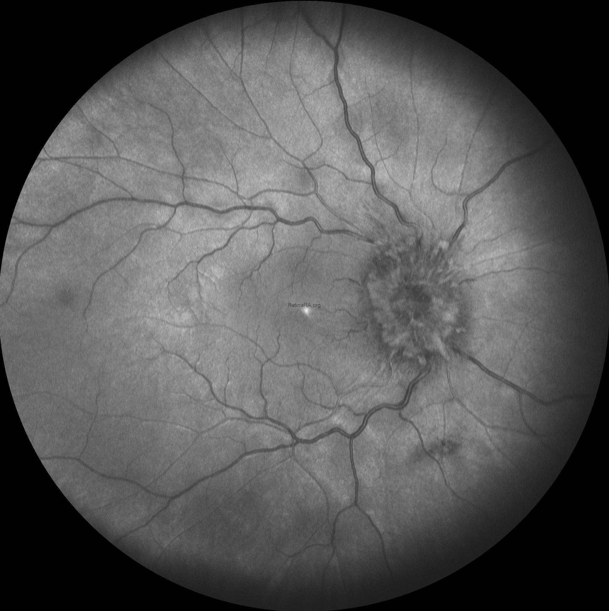
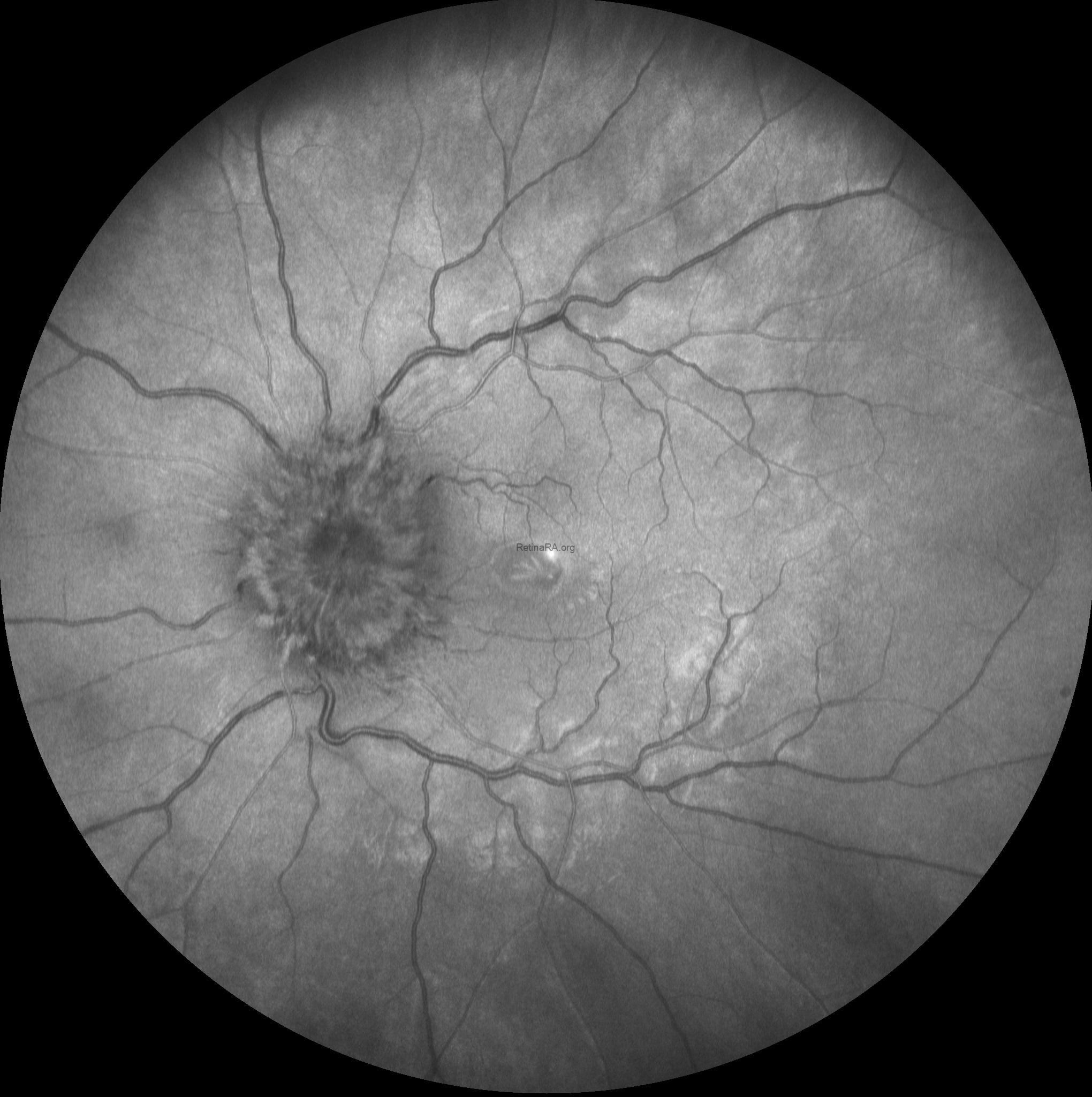
Based on the optic disc appearence, the patient was diagnosed as severe papilledema and consulted to the neurology department. After neurological asessment including craniyal computed tomography the patient was diagnosed as glioblastoma multiforme.
Papilledema refers to optic disc swelling secondary to increased intracranial pressure. It is typically bilateral and may present with symptoms such as headache, transient visual obscurations, nausea, and vomiting. On fundoscopic examination, the optic disc appears elevated with blurred margins, hyperemia, and sometimes hemorrhages or cotton wool spots. Papilledema is a clinical emergency, as it may indicate serious underlying conditions such as brain tumors, cerebral venous sinus thrombosis, or idiopathic intracranial hypertension. Prompt diagnosis and management are essential to prevent permanent vision loss.
