This was a 42-year old female admitted to outpatient clinic for routine eye check-up. The patient had no known systemic disease and was not on any systemic or ocular drugs. Family history of the patient was also unremarkable. The BCVAs were 20/20 for both eyes and IOPs were within normal limits. Anterior segment examinations were also normal.
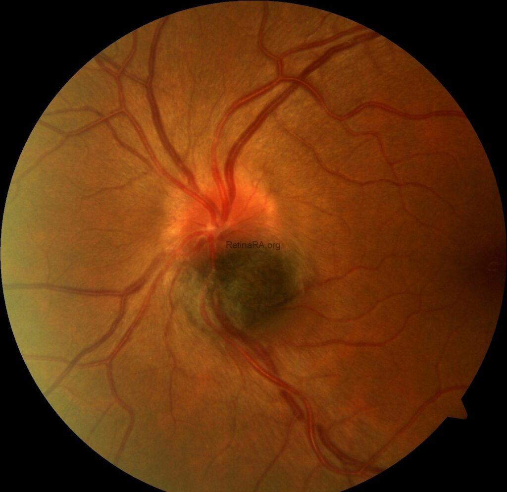
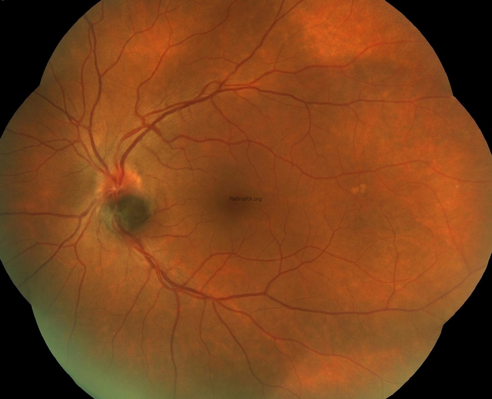
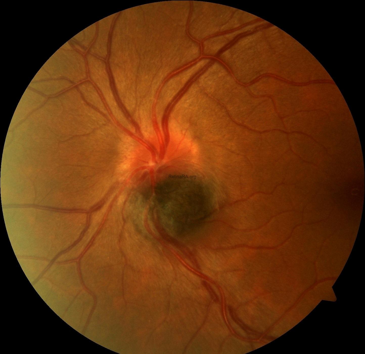
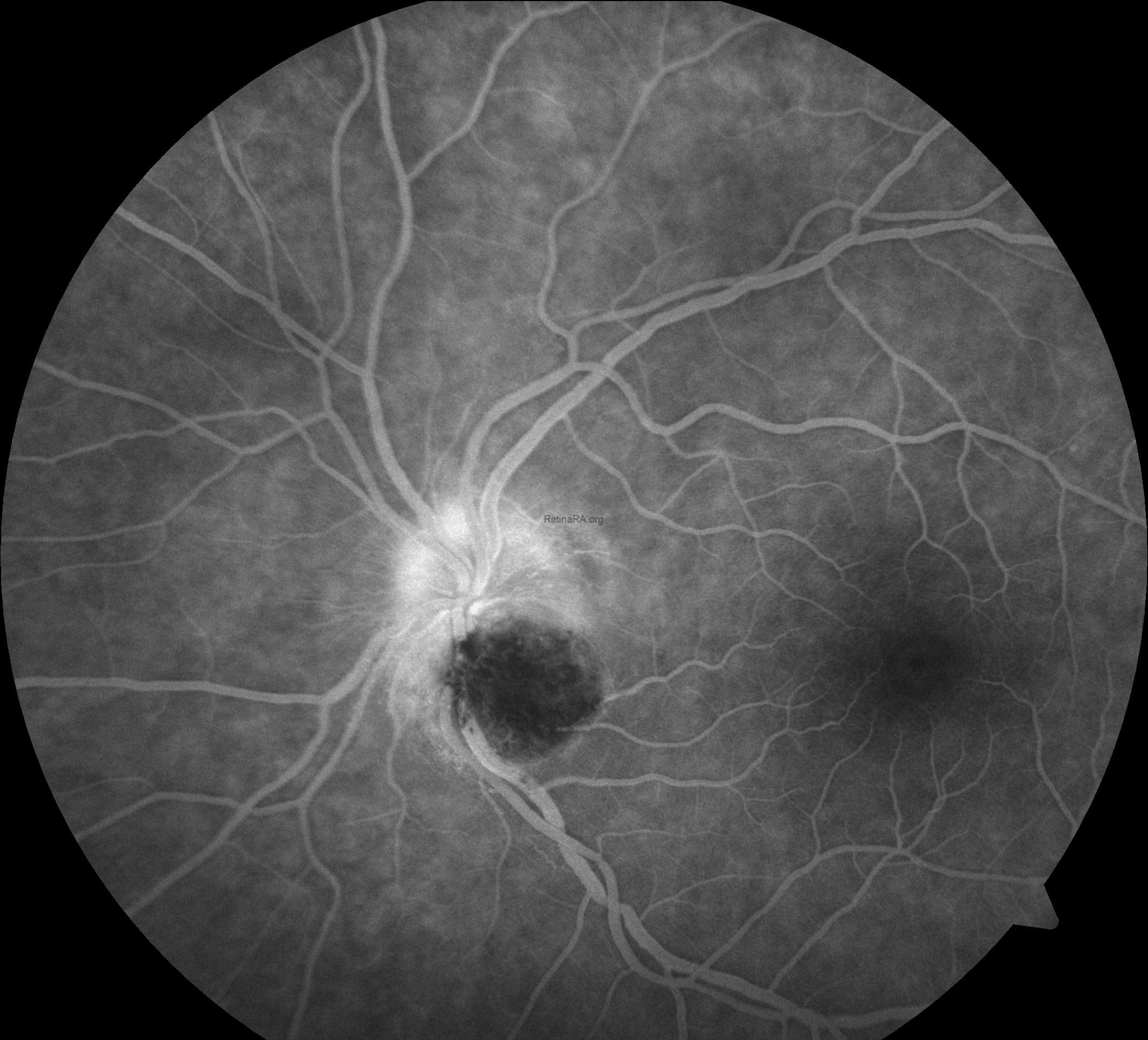
While the fundus examination of the right eye was completely normal; the left eye showed well-defined gray-black pigmentation at the inferior margin of the optic disc extending to the inferiortemporal retina. The lesion was elevated with feathery margins about 1.0 disc-area in size with the involvement of adjacent choroid. Lipofuscin deposition, retinal hemorrhages, retinal edema and subretinal fluid were absent. There were not any other retinal lesions or ocular melanocytosis. The vitreous was clear. Pupillary light reflexes were brisk and equal bilaterally.
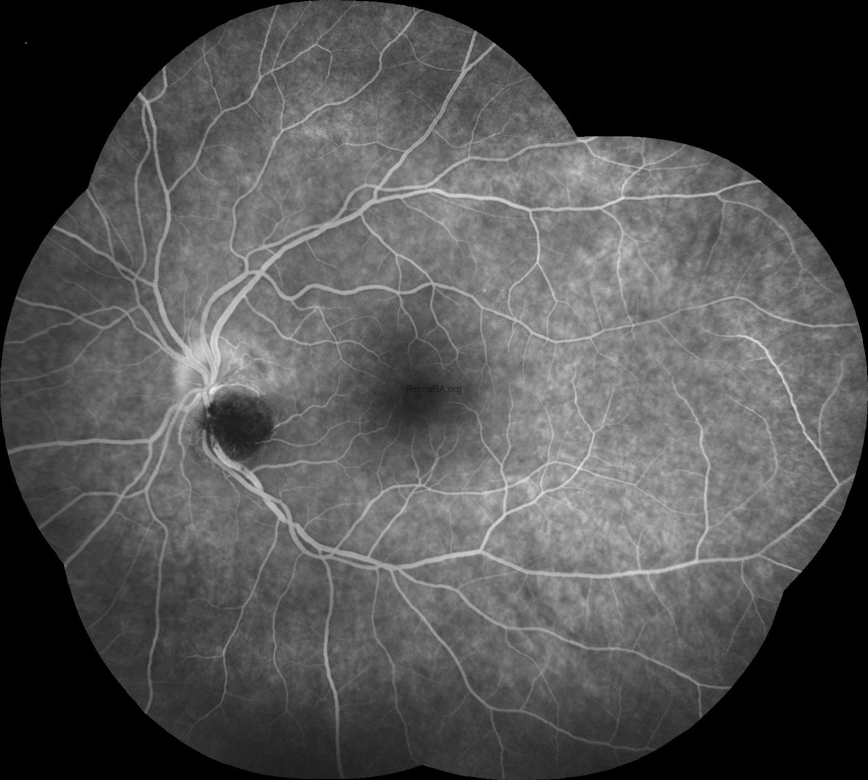
Fundus fluorescein angiography revealed hypo-fluorescence of the mass lesion in all stages without any evidence of late phase hyper-fluorescence. There was staining of the disc margins superiorly.
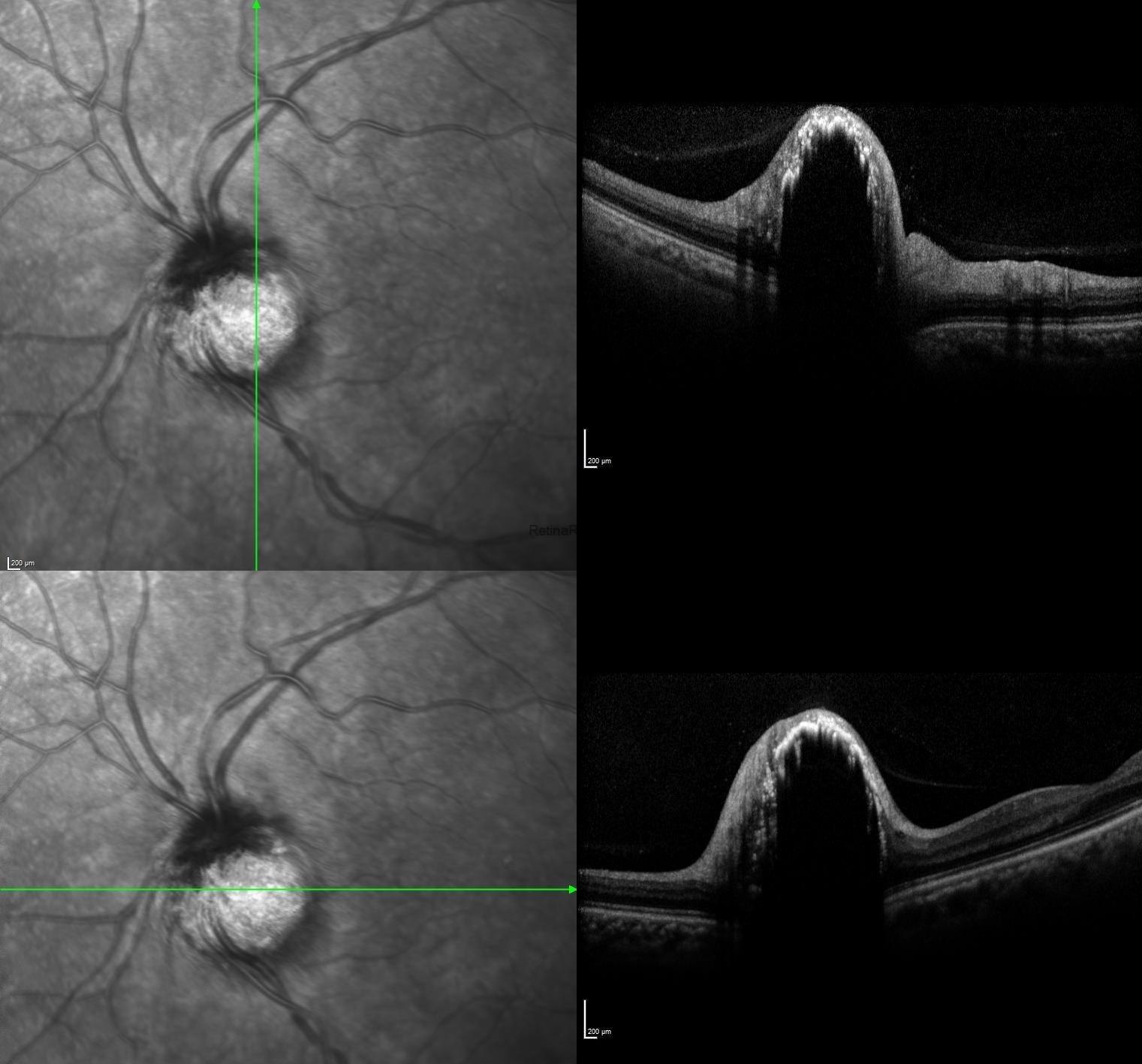
Optical coherence tomography scans passing through the lesion exhibited nodular elevated lesion with areas of irregular hyperreflectivity overlaying the tumor in additon to dense posterior shadowing effect.
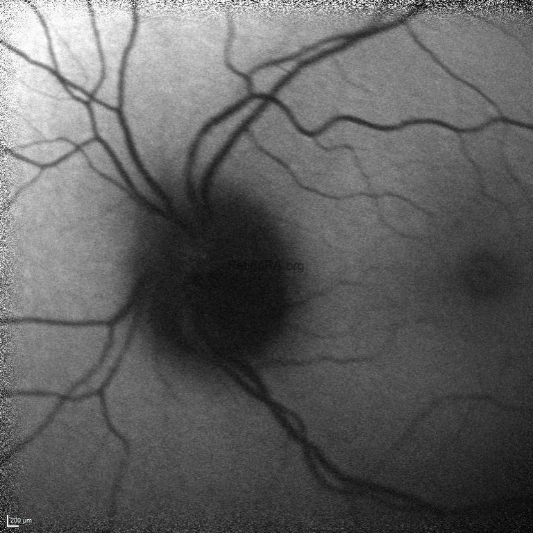
Fundus autofluoresce demonstrated hypo-autofluorescence corresponding to the pigmented mass on the optic disk.
Melanocytoma of the optic disc is a static tumor often found in an incidental routine ophthalmic assessment. These lesions are frequently unilateral with a little preference to affect visual acuity. Regarding the clinical presentation and vision, life-threatening melanoma represents a critical differential diagnostic challenge because of its subtle clinical similarities with melanocytoma. Thus, it is important to differentiate melanocytoma from malignant melanoma. By using multimodal imaging modalities, such as fundus photography, short-wave autofluorescence, fluorescein angiography, and optical coherence tomography, this discrimination is possible. Different from the melanosctoma; malignant melanoma is characterized by a vascular mass lesion with orange pigmentation suggestive of lipofuscin, which is hyper-autofluorescent as well as it is usually more than 2 mm in thickness on echography and may show vascularization and serous detachment.
Credit: Kemal Tekin, M.D., from Ulucanlar Eye Training and Research Hospital
Instagram accounts: @retina.academy and @dr.kemaltekin
