A 56-year-old female patient with retinal neovascularization (neovascularization elsewhere – NVE) secondary to Type 2 DM.
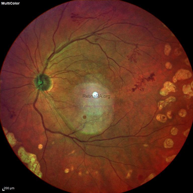
In the multicolor image, NVEs were seen as tangle-shaped new vessel formations (yellow rings) originating from the retinal vessels. Additionally, laser photocoagulation scars could be observed in the multicolor image.
Multicolor imaging of retinal neovascularization elsewhere (NVE)

In the OCT scans passing over the NVEs, these new vessels were observed as hyperreflective formations extending from the retinal surface to the vitreous. Observing the extension to the vitreous on OCT allows these vessels to be distinguished from IRMA and collateral vessels.
OCT of retinal neovascularization elsewhere (NVE)


On ultra-widefield fluorescein angiography, NVEs were seen as leaky vessels. In addition, the borders of pan-retinal laser photocoagulation were more clearly defined. In this patient, laser photocoagulation should be advanced to the macular border for regression of NVEs.
Ultra-widefield fluorescein angiography of retinal neovascularization elsewhere (NVE)

Credit: M. Giray Ersoz, MD, FEBO
Biruni University School of Medicine, Department of Ophthalmology, Istanbul, Turkey
Instagram accounts: @retina.review and @retina.dr.girayersoz
