
This was a 56-year-old female without any systemic disease who applied to emergency department with decreased vision in her left eye. The patient had been follow-up by glaucoma department due to chronic angle closure glaucoma and undergone laser iridotomy before. The BCVAs were 20/20 in the right eye and counting fingers in the left eye while the IOPs were measured as 15 mmHg and 19 mmHg, respectively for the right and left eyes.
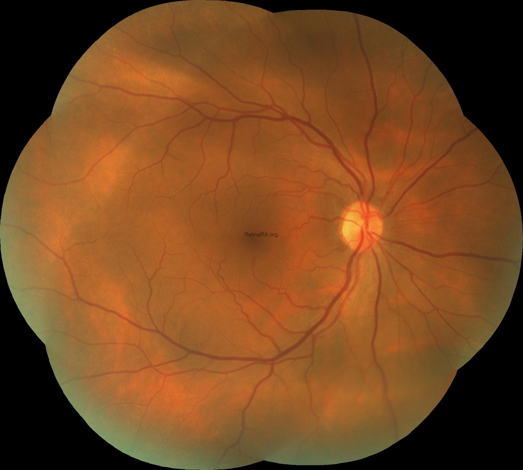
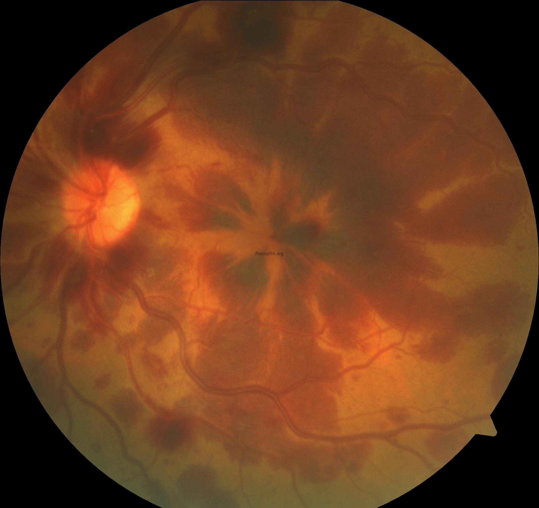
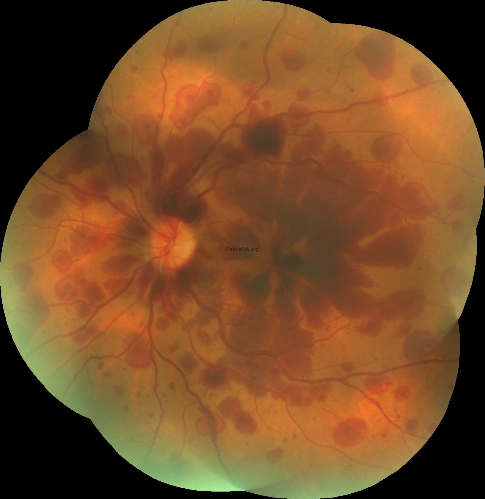
Fundus examination of the left eye demonstrated scattered petaloid-shaped, deep retinal haemorrhages radiating from the fovea and mildly dilated and tortuous retinal veins, consistent with central retinal vein occlusion in the left eye; while the right eye was completely normal.
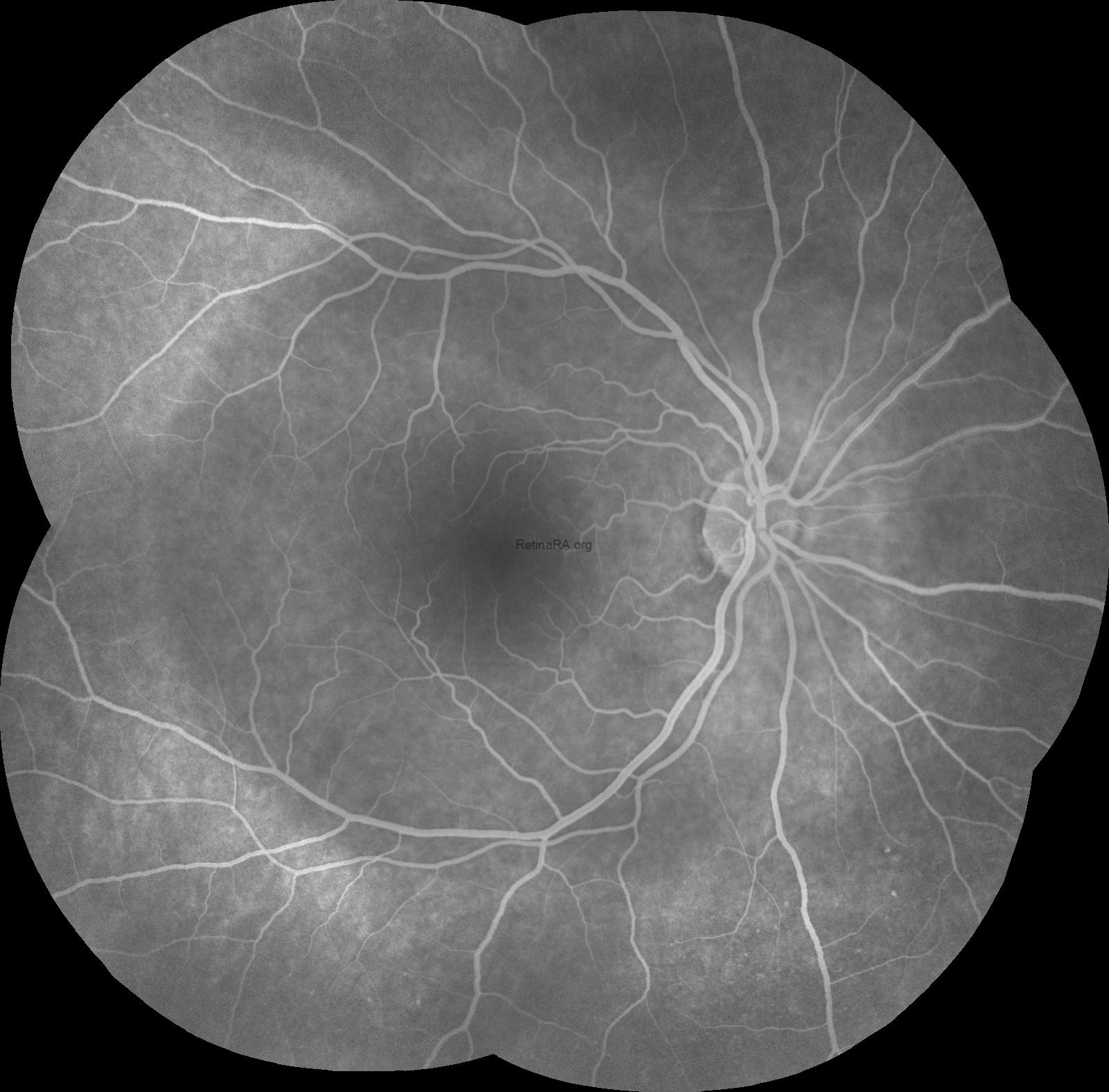
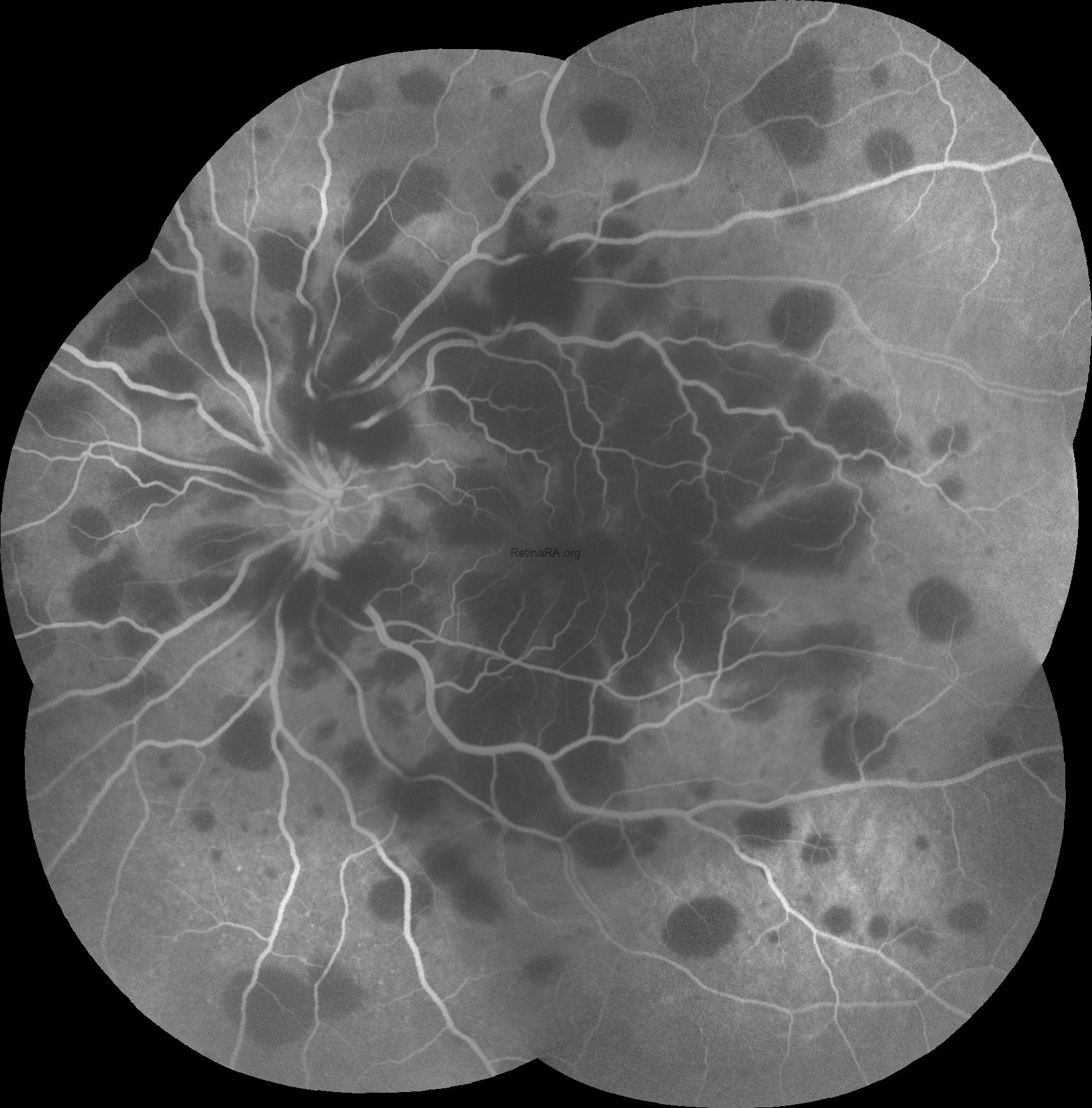
Fluorescein angiography revealed areas of blocked fluorescence corresponding to retinal haemorrhages without any leakage in the left eye, while there were not any pathology in the right eye.
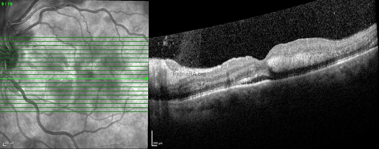
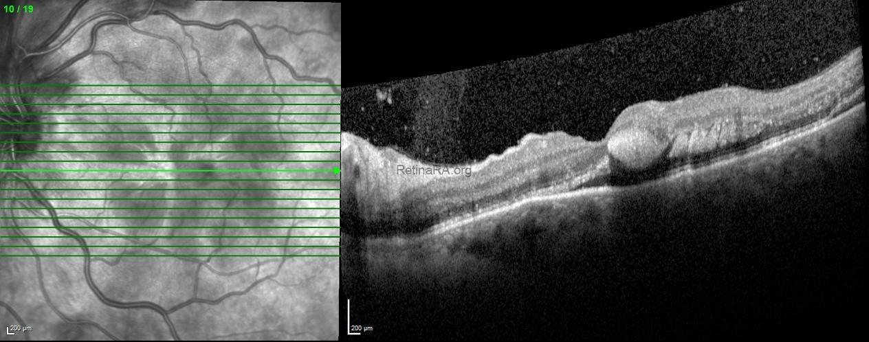
Spectral-domain optical coherence tomography of the left eye showed a central foveal hemorrhage and adjacent hemorrhages radiating in the Henle fiber layer in addition to mild serous retinal detachment.
A systemic work up by internal medicine, cardiology, hematology and nephrology departments was performed for the patient and no pathology was detected.
Henle fibre layer haemorrhage typically presents with a petaloid pattern of haemorrhage located in the henle fiber layer of the retina. Optical coherence tomography findings of blood located within the Henle fiber layer were described as characteristic hyperreflectivity from the hemorrhage delineated by the obliquely oriented fibers in the Henle layer. The deep capillary plexus of the retina was recently reported to be the predominant part of the retina with respect to venous outflow that indicates a possible reason for the location of haemorrhages in the Henle layer. HFLH can be associated with a variety of ocular and systemic conditions such as increased systemic central venous pressure (intracranial haemorrhage caused by trauma or aneurysm, trauma, general or epidur alanaesthesia, Valsalva retinopathy, and hypertension), local retinal venous pressure (branch or central retinal vein occlusion, decompression retinopathy, and local ocular trauma), or local pathologies affecting the deep capillary plexus (polypoidal choroidal vasculopathy, macular telangiectasia type 2, myopic degeneration, and age-related macular degeneration).
Credit: Kemal Tekin, M.D., from Ulucanlar Eye Training and Research Hospital
Instagram accounts: @retina.academy and @dr.kemaltekin