A 62-year-old female presented with decreased and blurry vision in her left eye. Visual acuity was 0.6 in the left eye and 1.0 in the right eye. Anterior segment examination was unremarkable in both eyes.
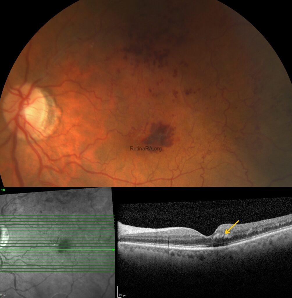
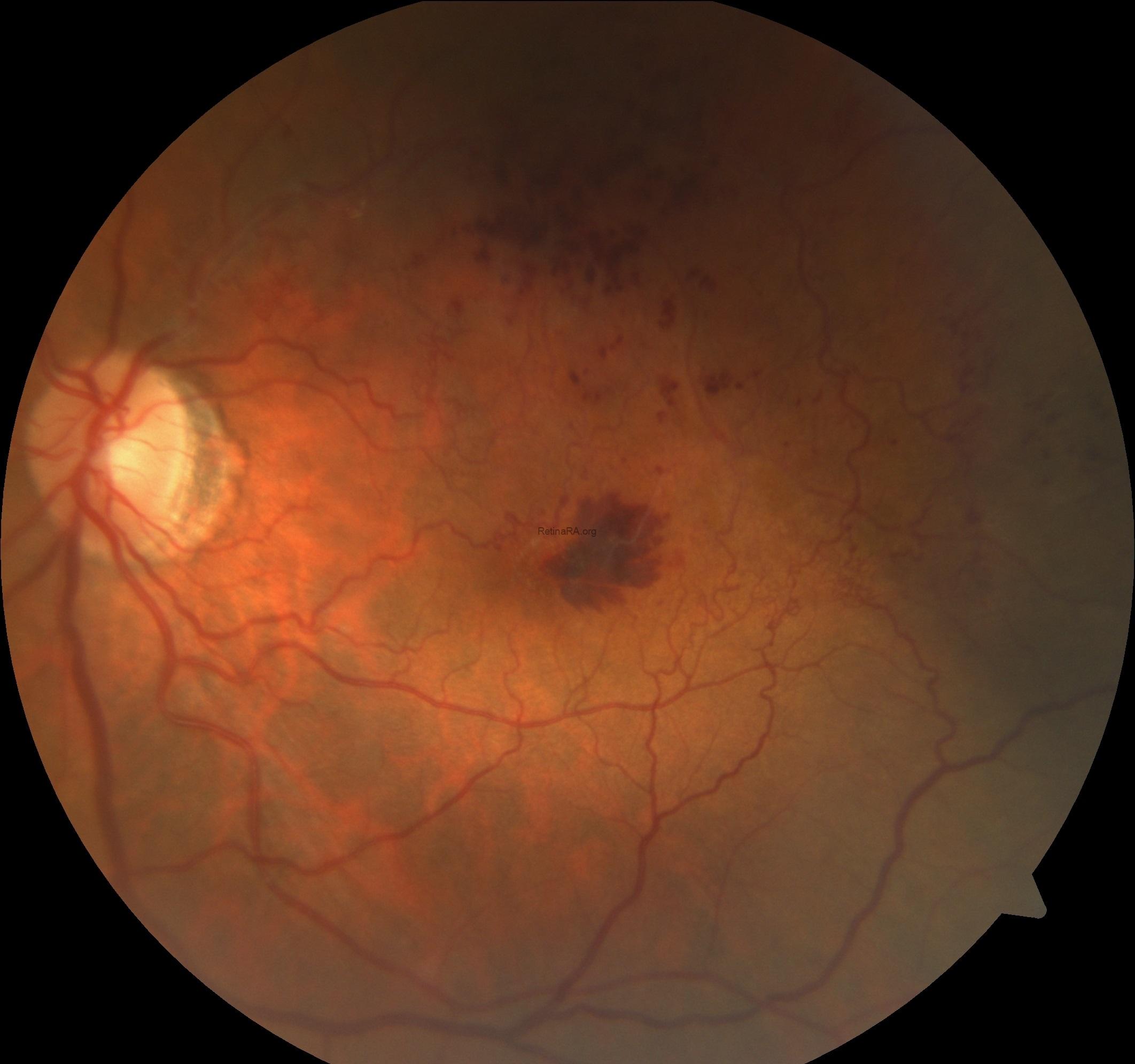
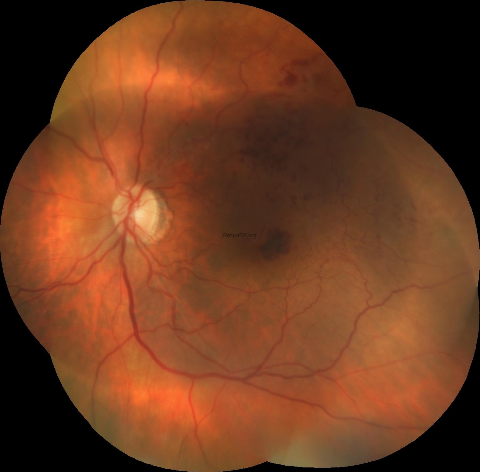
Fundus examination of the left eye revealed feathery (tufy) hemorrhages in the temporal foveal region, accompanied by dot-blot hemorrhages and prominent collateral vessels in the superior and temporal macula.
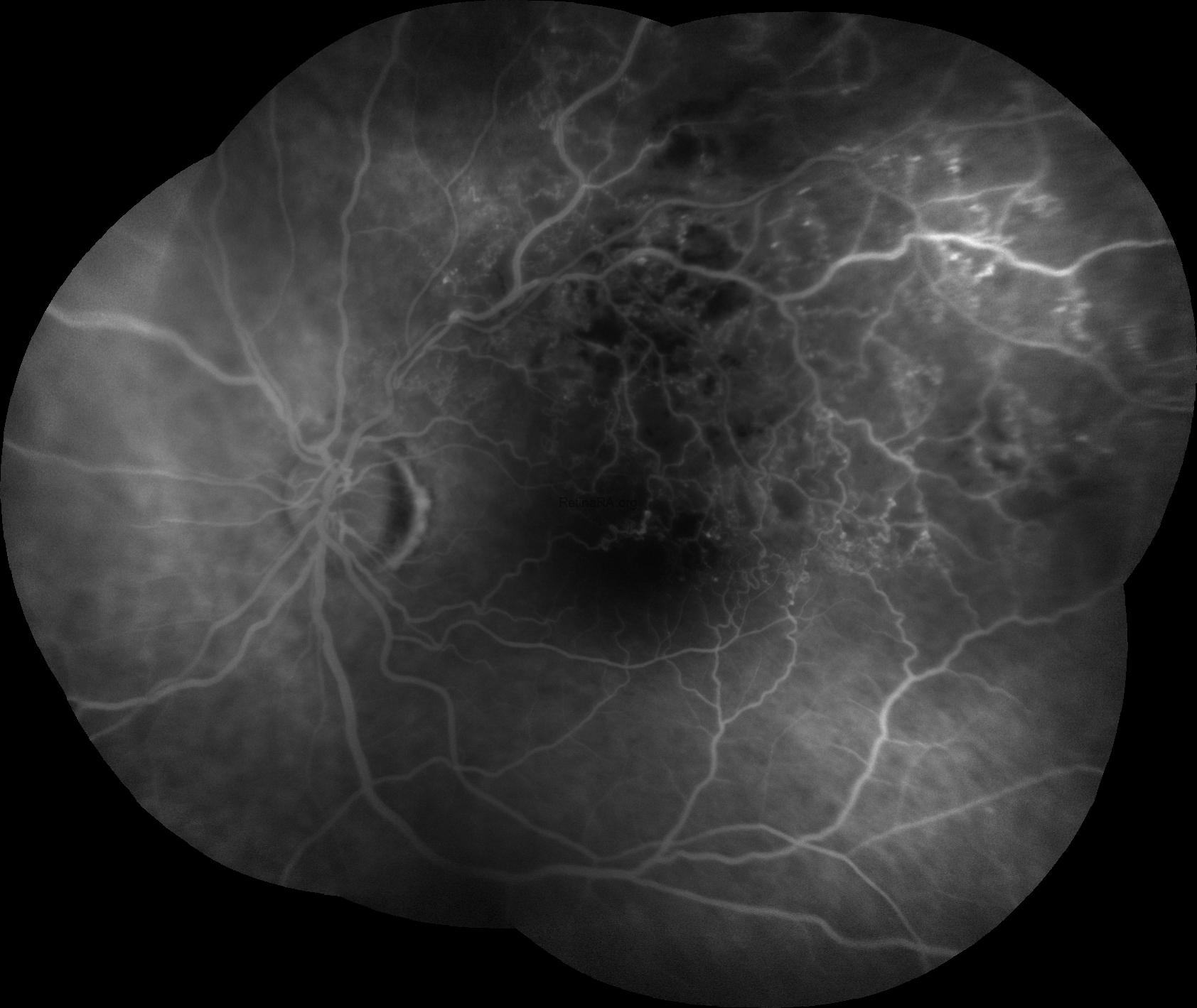
Fluorescein angiography revealed areas of retinal ischemia and confirmed the presence of collateral vessels in the superior and temporal macula.
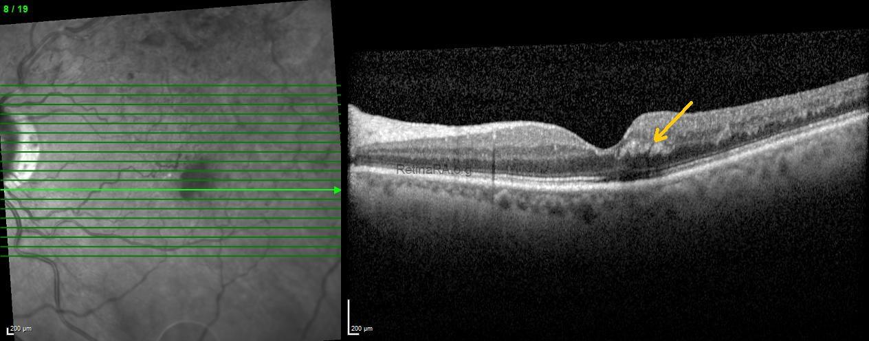
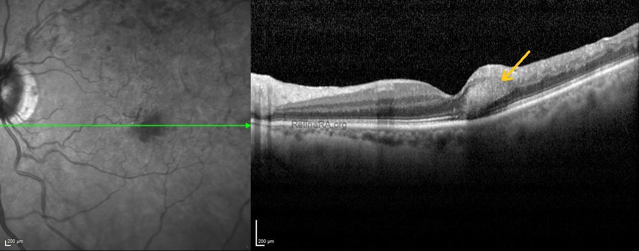
Optical coherence tomography scans passing through the feathery hemorrhages revealed obliquely oriented hemorrhages with shadowing within the Henle fiber layer at the temporal fovea (indicated by arrows).
These findings were consistent with a Henle fiber layer hemorrhage associated with branch retinal vein occlusion. The patient was monitored for spontaneous resolution, with follow-up OCT and FFA planned to assess ischemia and collateral vessel progression.
Henle fiber layer hemorrhage is a distinct type of deep intraretinal hemorrhage localized in the macula, following the oblique orientation of Henle fibers. On fundus examination, it often appears feathery or petaloid, radiating from the fovea, while OCT shows hyperreflective lesions with shadowing in the Henle fiber layer. These hemorrhages are usually associated with vascular disturbances in the deep capillary plexus. Although they can occur in various retinal vascular conditions, Henle fiber layer hemorrhages may also be seen in eyes with branch or central retinal vein occlusion. Recognizing this pattern is important for accurate diagnosis and management.
Credit: Kemal Tekin, M.D., from Ulucanlar Eye Training and Research Hospital
Instagram accounts: @retina.academy and @dr.kemaltekin
