Patient history
A 34-year-old male patient with a history of chronic central serous chorioretinopathy (CSC) in both eyes since 2020 presented for follow-up. The patient works in the construction sector and has a history of allergic rhinitis treated with intranasal corticosteroids.
Best-corrected visual acuity (Snellen) was 20/50 in the right eye (OD) and 20/800 in the left eye (OS).Multimodal imaging was performed, including ultra-widefield fundus photography, ultra-widefield autofluorescence, spectral-domain OCT, OCT angiography, and TowardPi imaging in both eyes
Images & descriptions
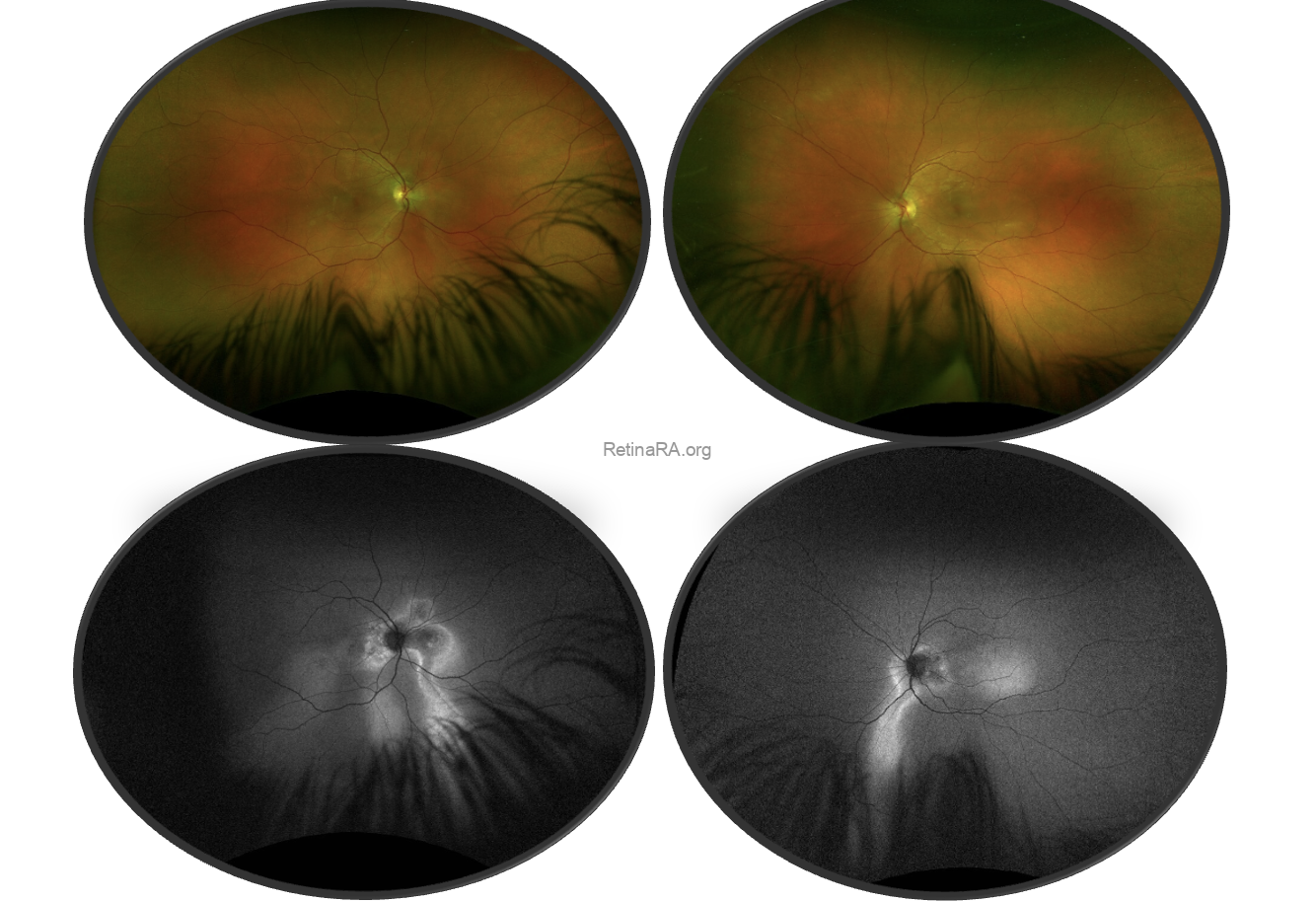
Ultra-widefield fundus photography showed extensive retinal pigment epithelium (RPE) alterations with gravitational tracks in both eyes, consistent with chronic CSC.
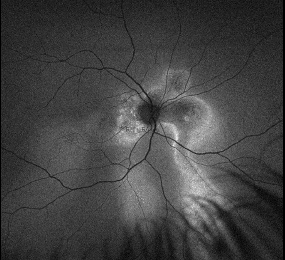
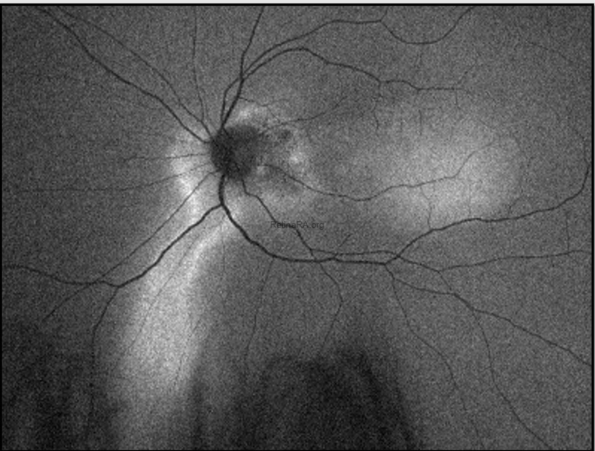
Ultra-widefield autofluorescence revealed hypoautofluorescent gravitational tracks corresponding to areas of RPE atrophy, surrounded by hyperautofluorescent borders indicating ongoing RPE dysfunction.
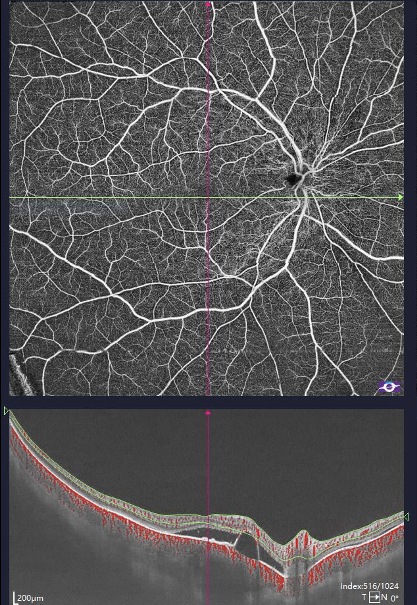
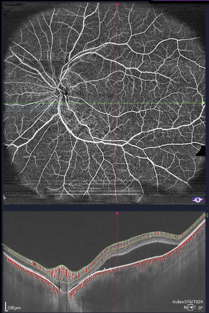
Spectral-domain OCT showed irregular RPE, areas of RPE atrophy, pachychoroid features, and chronic structural changes. No obvious active subretinal fluid was detected, but there were diffuse RPE alterations consistent with chronic disease.
OCT angiography did not reveal any macular neovascularization. The superficial capillary plexus was preserved, while the choriocapillaris slab showed flow signal voids corresponding to areas of RPE alteration, consistent with chronic CSC
Discussion
Chronic central serous chorioretinopathy is part of the pachychoroid disease spectrum and is characterized by choroidal thickening, RPE alterations, and gravitational tracks due to chronic subretinal fluid displacement. Over time, chronic RPE decompensation can lead to diffuse retinal pigment epitheliopathy and significant visual loss.
This case illustrates the typical multimodal imaging features of chronic CSC, including gravitational tracks on fundus photography, hypo- and hyperautofluorescent changes on autofluorescence, pachychoroid features on OCT, and choriocapillaris alterations on OCT angiography. Multimodal imaging is essential to assess disease chronicity, detect complications such as macular neovascularization, and guide management.
Affiliation
Safa Chaabane, MD
Retina Department, Intercommunal Hospital Center of Creteil, France
Head of department: Prof. Eric Souied
Instagram: @safachaabane_ | @creteilretinateam