A 70-year-old female who has been followed-up due to age-related macular degeneration for approximately 10 years presented with decreased vision in her left eye that was realized 1-month ago with visual acuity of 20/200. Colored fundus image revealed wide-spread soft drusen, aggregation of confluent drusen, and central macular hole with pigment epithelial detachment.

Macular hole and drusen were seen as hyper-autofluorescence in fundus autofluorescence.

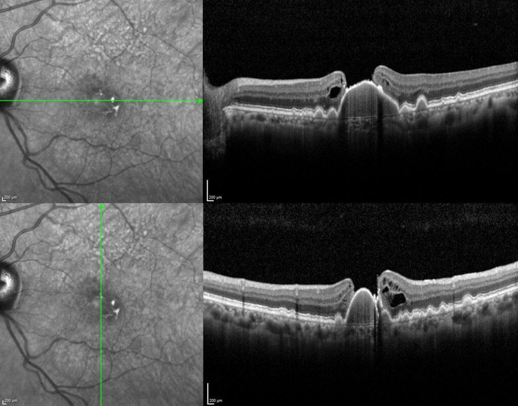
The horizontal and vertical optical coherence tomography scans passed through the central fovea revealed sub-RPE deposits, a large drusenoid pigment epithelial detachment combined with full-thickness macular hole with a detached posterior hyaloid.

Full-thickness macular hole (FTMH) associated with pigment epithelial detachment (PED) is a very rare entity. A few cases of a FTMH developing on a serous PED have been reported. Pathologic mechanisms and treatment outcomes are uncertain. In regard to the pathogenesis of FTMH development in eyes with drusenoid PEDs, it has been proposed that different mechanical forces were involved in the pathogenesis: in addition to anteroposterior and/or tangential traction at the vitreomacular interface, a retracting force within PED owing to flattening of drusenoid PED, or a force pushing out of the drusenoid PED as it grew, may have played a role.
Credit: Kemal Tekin, M.D., from Ulucanlar Eye Training and Research Hospital
Instagram accounts: @retina.academy and @dr.kemaltekin