
This patient was a 32-year-old male who was referred for evaluation of macular degeneration. The patient has been follow-up by nephrology department of another clinic and underwent two prior renal transplantation due to glomerulonephritis, last was 5 years ago. The BCVAs were 1/10 for both eyes and anterior segment examination showed no abnormality.
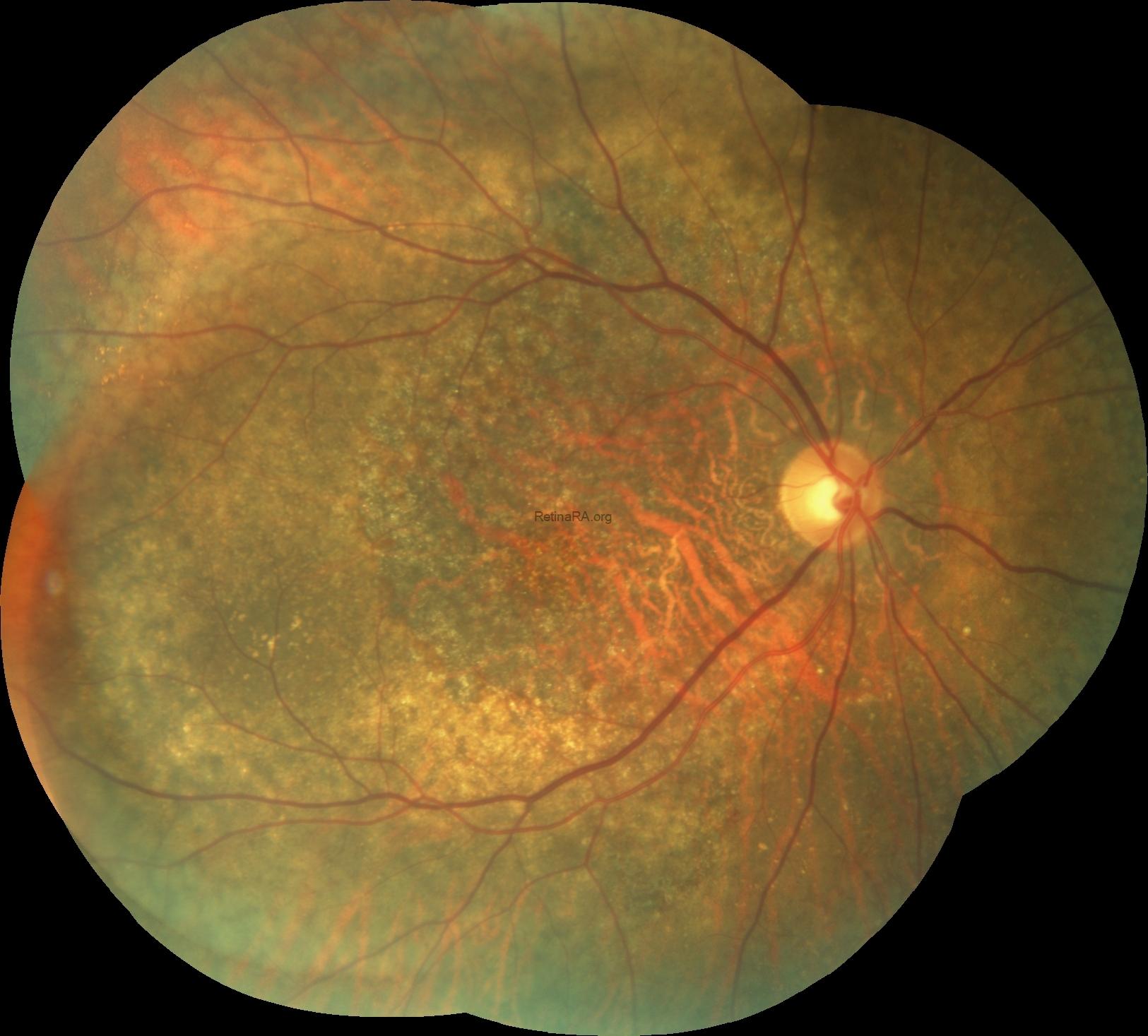
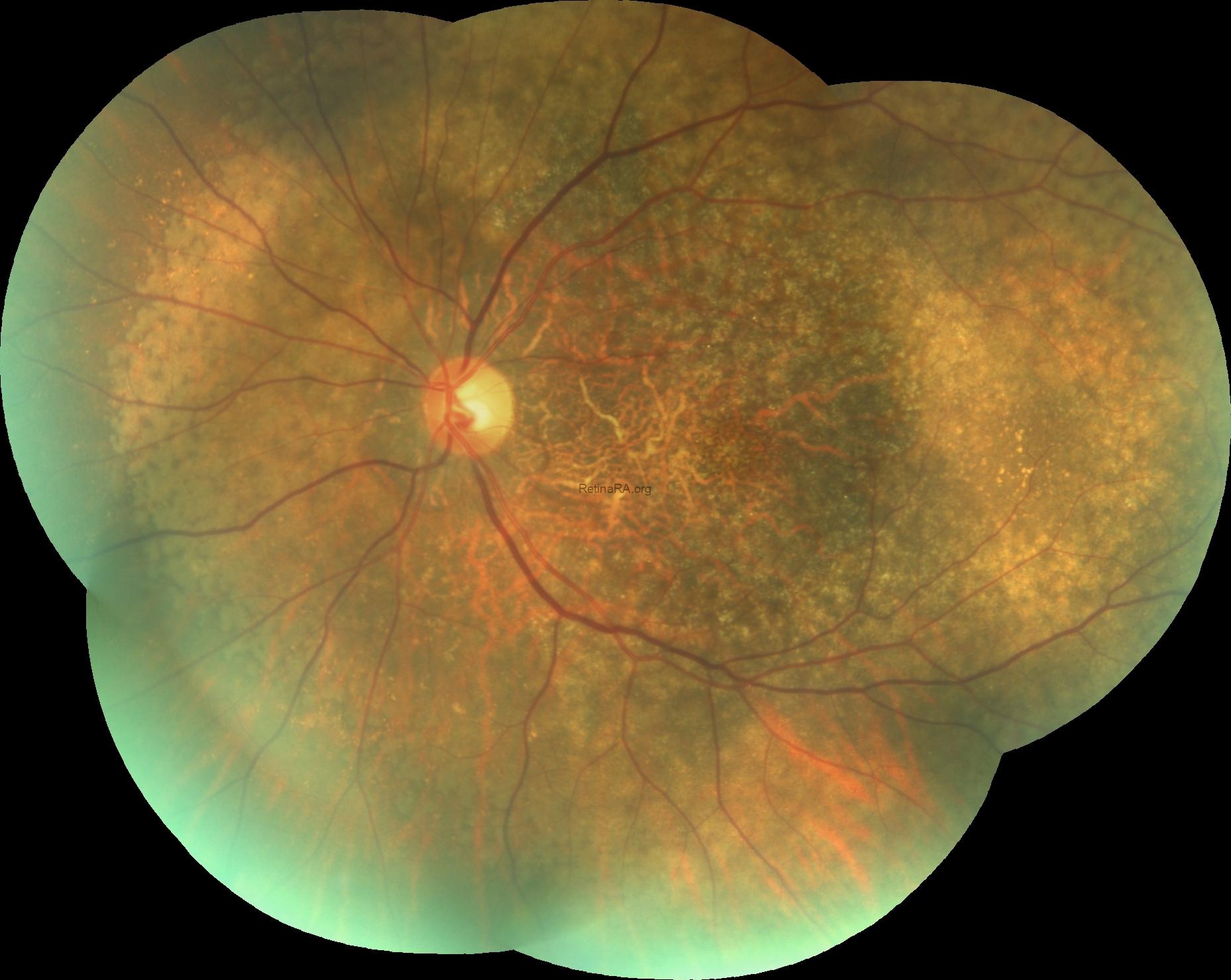
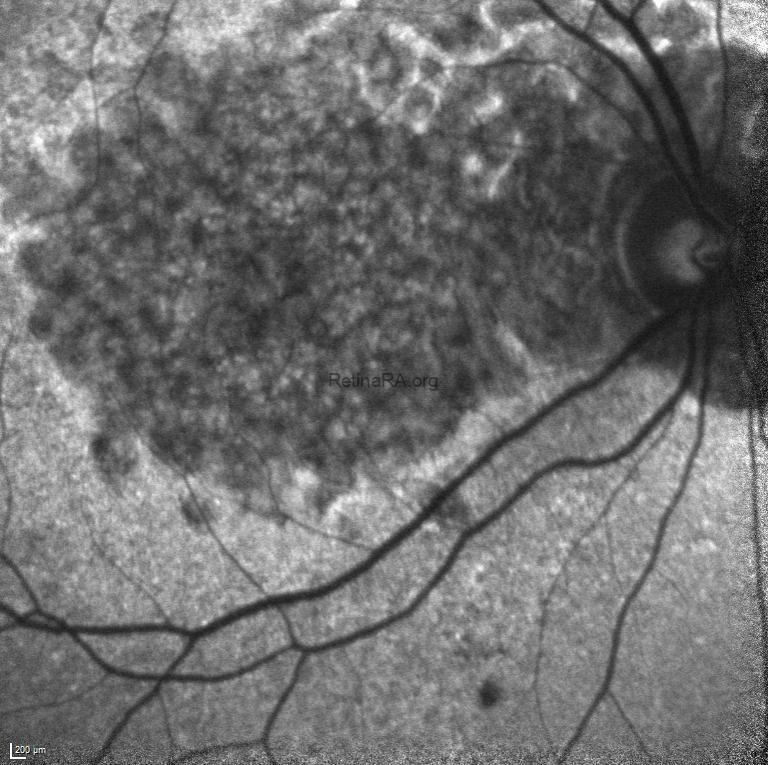
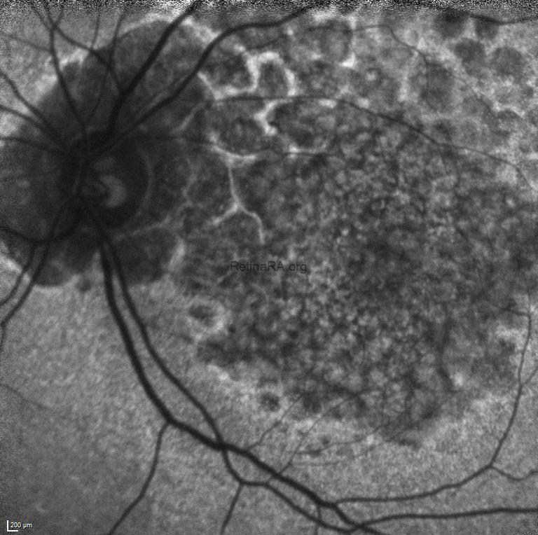
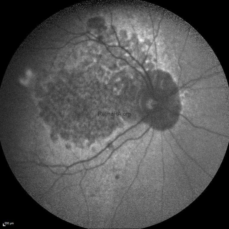
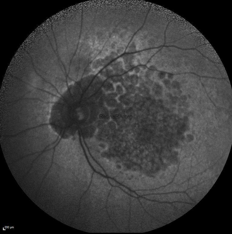
Fundus examination of the right and left eyes revealed subretinal pigment epithelium drusen-like deposits extending from the macula to the mid periphery in both eyes in addition to macular atrophy.
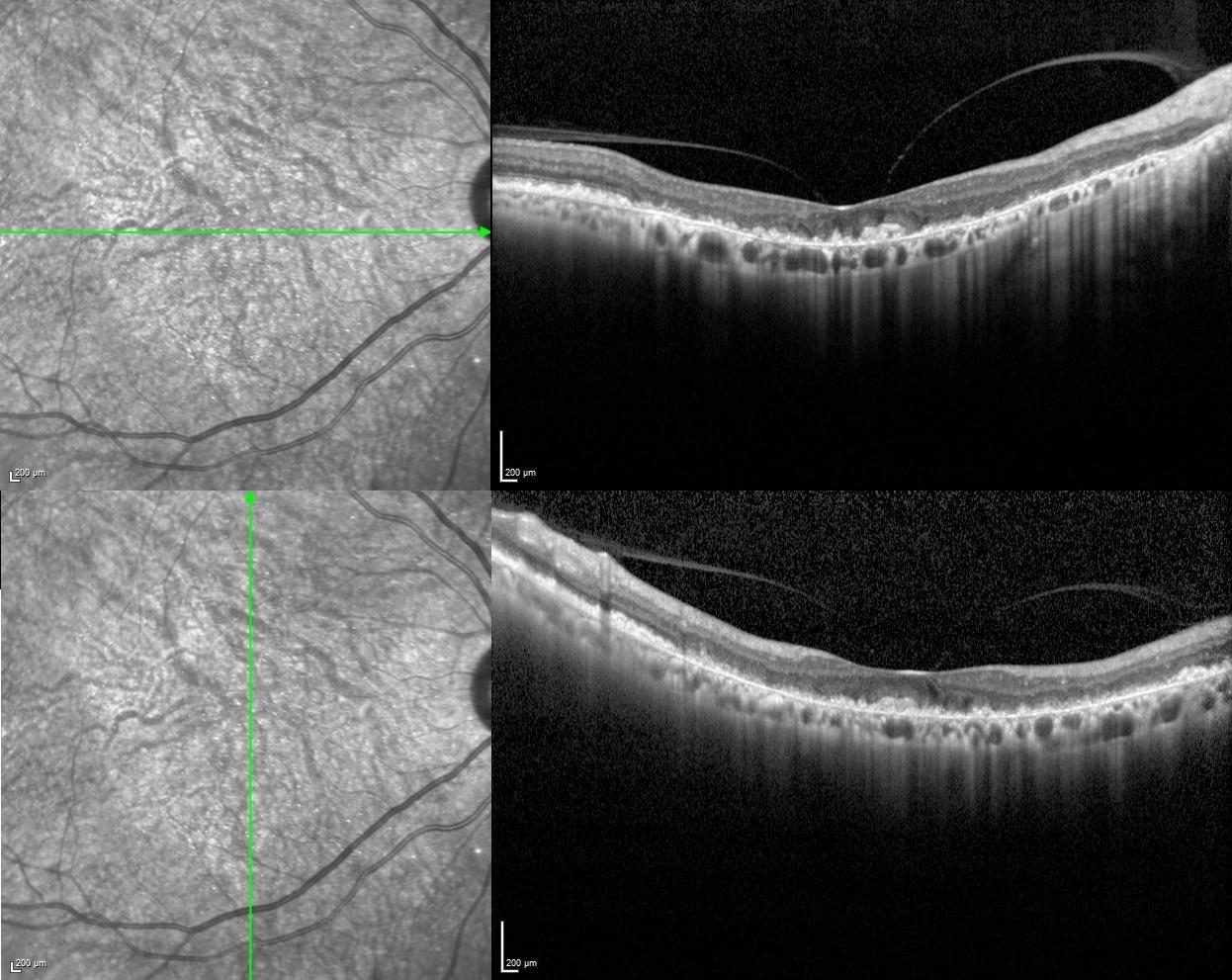
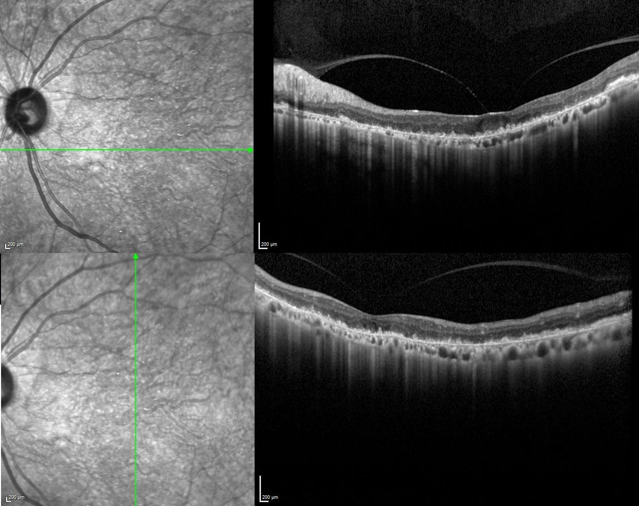
Vertical and horizontal optical coherence tomography scans revealed subretinal accumulation of drusen-like deposit in additon to outer retinal and choriocapillaris atrophy in both eyes.
Blue-light macular fundus autofluorescence and wide-field fundus autofluorescence revealed decreased autofluorescence at the macula and increased autofluorescence in mid-peripheral retina.
According to the medical history and multimodal imaging findings, the patient was diagnosed with diffuse drusen-like deposits associated with dense deposit disease, also known as C3 glomerulopathy or membranoproliferative glomerulonephritis type II.
Atypical causes of drusen should be considered in young patients presenting with drusen and renal complaints should be questioned in these patients.
Credit: Kemal Tekin, M.D., from Ulucanlar Eye Training and Research Hospital
Instagram accounts: @retina.academy and @dr.kemaltekin
