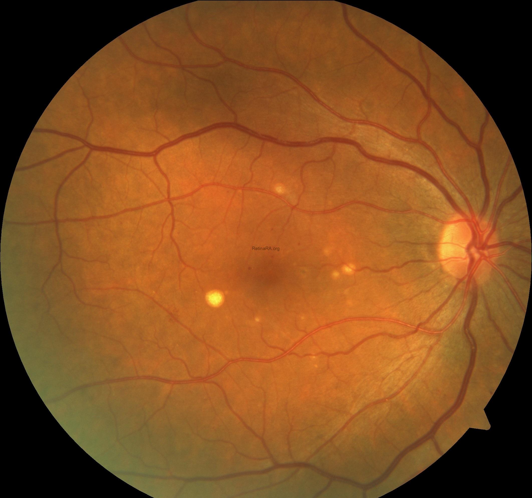
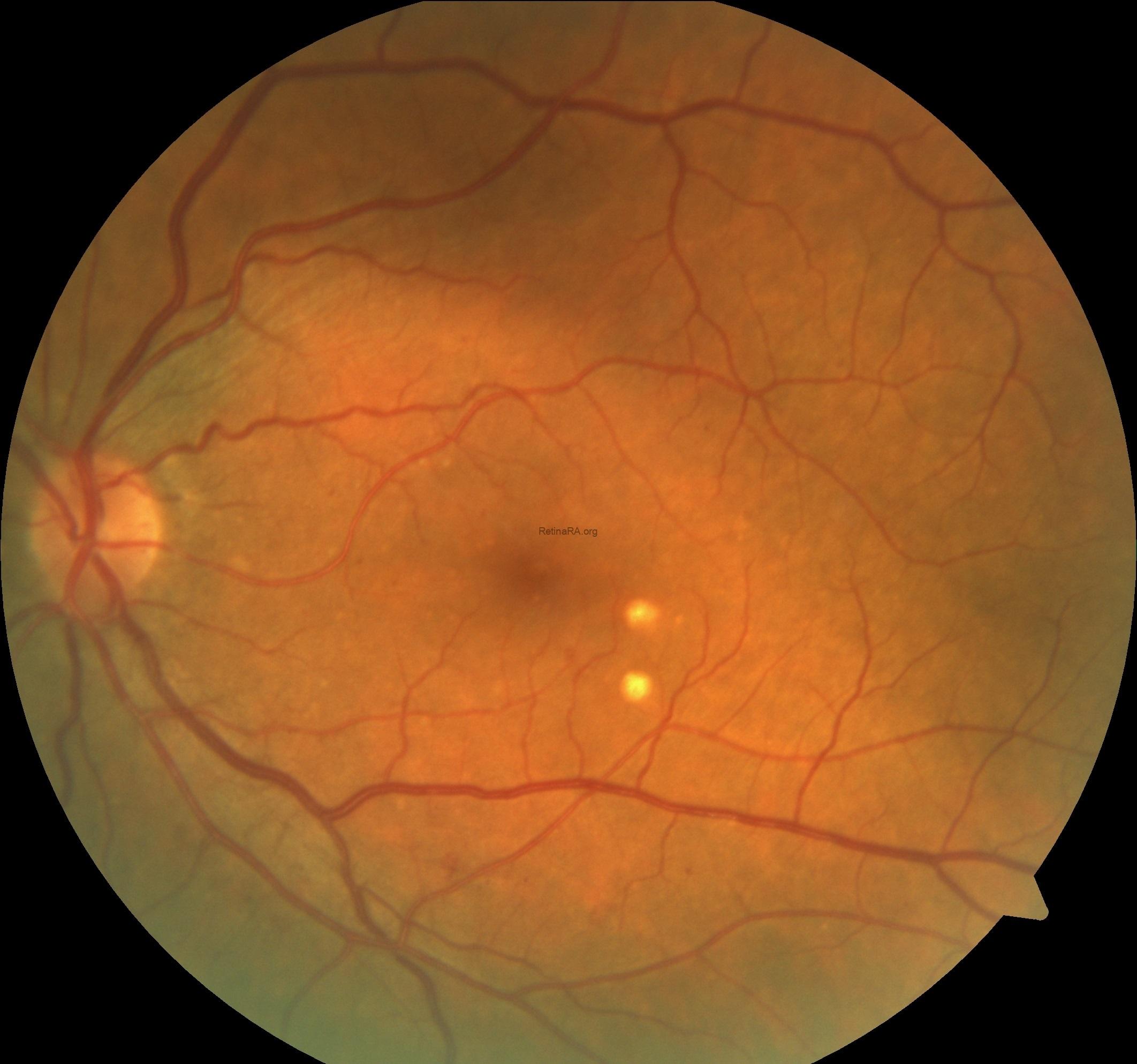
A 45-year-old female was referred to our retina clinic for evaluation of microaneurysms and yellowish deposits in both eyes. The patient’s visual acuities were 20/20 inboth eyes and anterior segment examinations were normal. She had type 2 diabetes mellitus and unremerkable ophthalmic history.
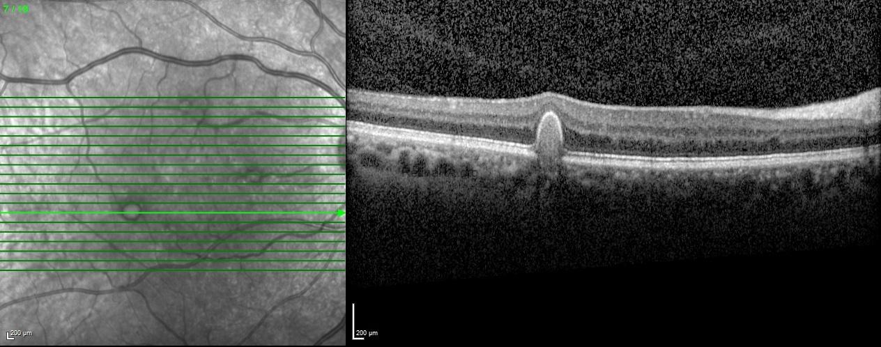
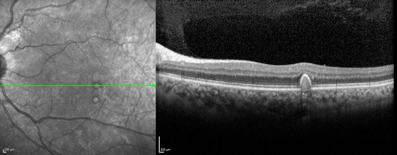
Fundus images of both eyes showed diabetic microaneurysms and a few yellow large drusen (
>125 μm) in the macula.
Spectral-domain optical tomography scans passing through the drusen demonstrated the dome-shaped retinal pigment epithelial elevations with variable internal reflectivity of drusen.
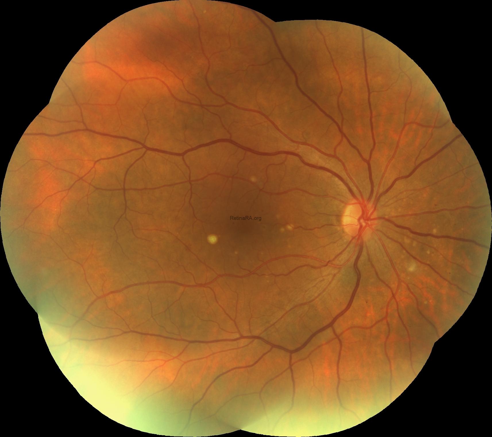
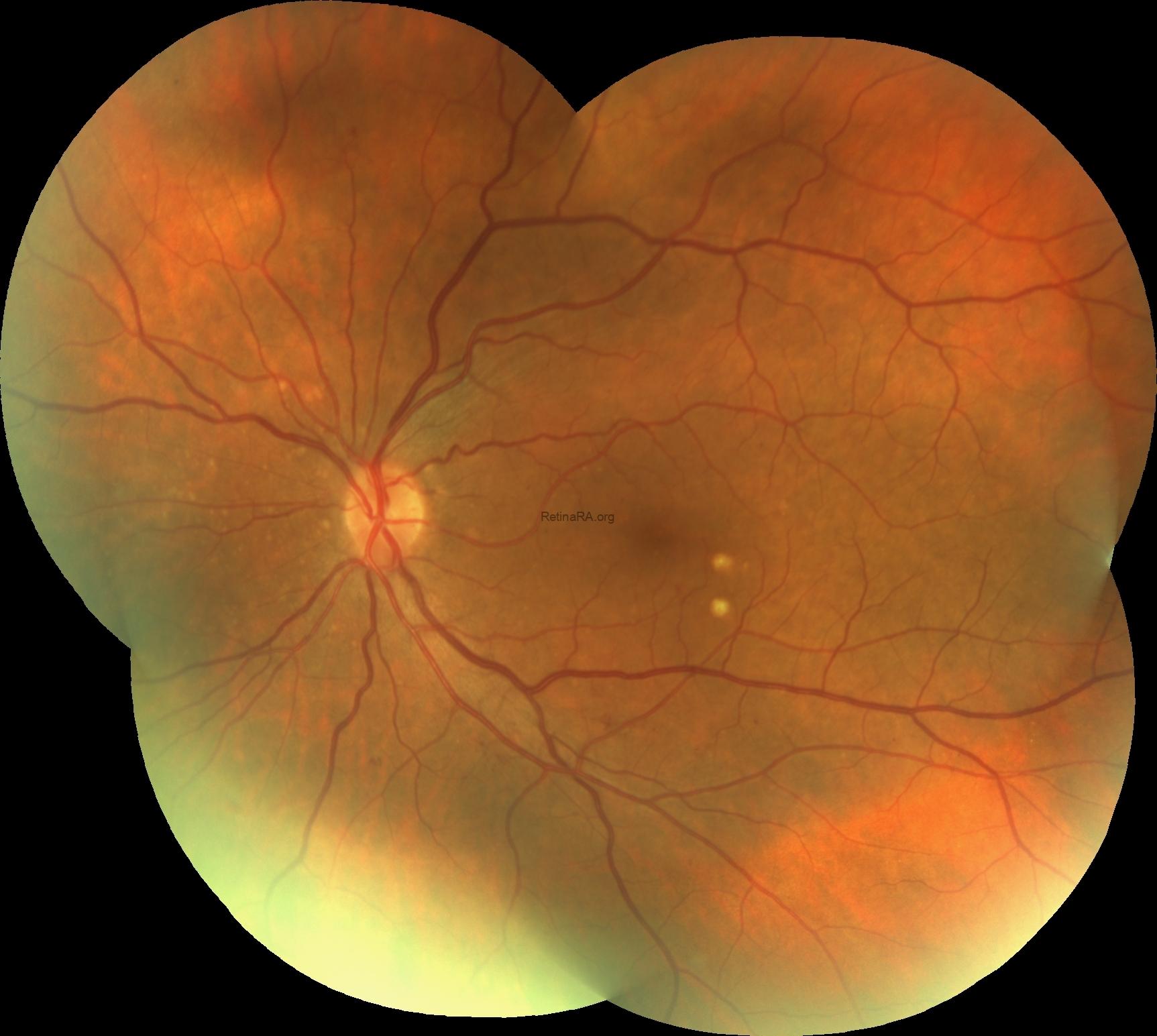
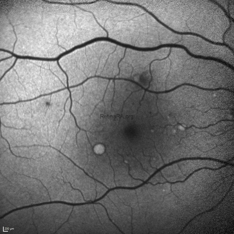
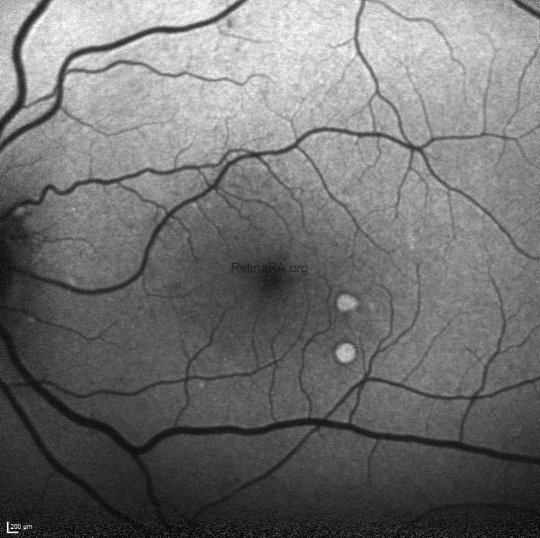
Fundus autofluorescence imaging exhibited the hyper-autofluorescence of drusen and hypo-autoflurescence of retinal microaneurysms and hemorrhages.
Drusen are extracellular deposits that accumulate between the retinal pigment epithelium and the inner collagenous layer of Bruch membrane. They are rarely observed in patients younger than 50 years. The commonly described types of early onset drusen are basal laminar drusen, malattia leventinese, and large colloid drusen (LCD), recently described. The origin and clinical significance of LCD remains unclear. Histologically, LCD are identical in composition and location to cuticular drusen, nodular drusen, or hard drusen.
Credit: Kemal Tekin, M.D., from Ulucanlar Eye Training and Research Hospital
Instagram accounts: @retina.academy and @dr.kemaltekin
