
This post will present a classical macular hole surgery.
This is a 62-year-old Male patient whose right vision has decreased for the last 2 months. He has a cataract and a macular hole in the right eye. The visual acuity of the right eye was 20/60.
Preoperative multicolor image, autofluorescence image, and OCT of the patient with macular hole
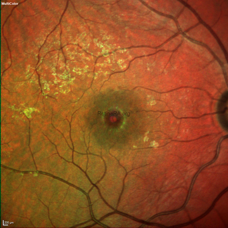


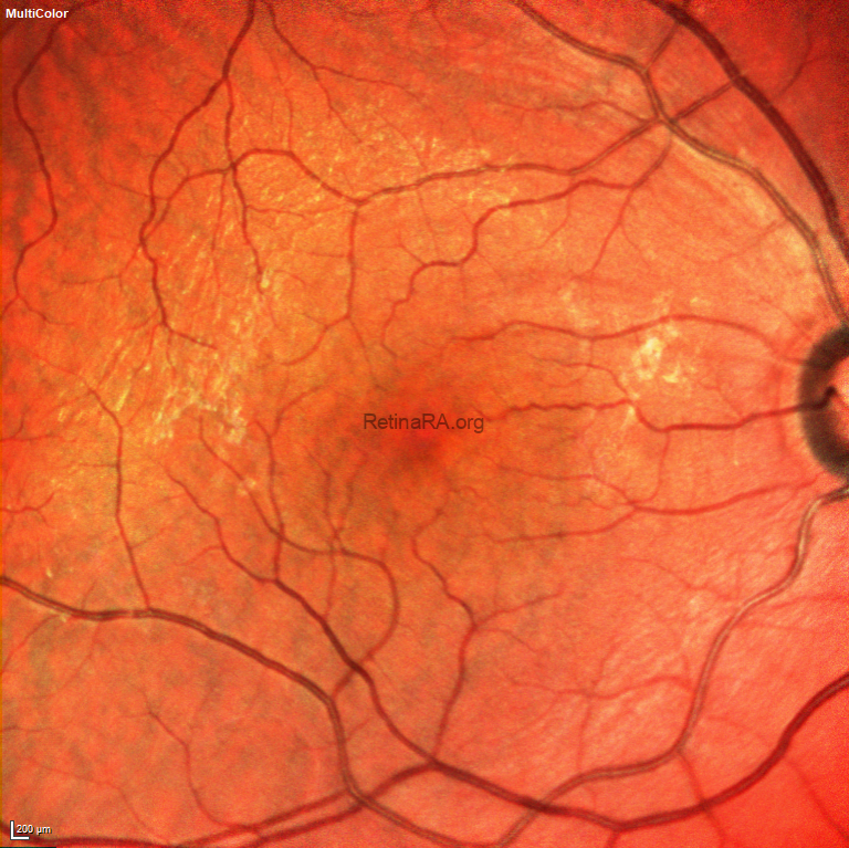
Various techniques have been described recently for large macular holes. However, the surgeon decided to perform classical macular hole surgery in this case because the hole was 220 microns wide. One month after the surgery, the hole had completely closed and visual acuity had increased to 20/20.
Postoperative multicolor image, autofluorescence image, and OCT of the patient with macular hole


Credit: Ayşegül Mavi Yıldız, MD, FEBO, FICO, MRCSEd
Retina Göz Hastanesi, Bursa, Türkiye
Instagram account: @aysegul_md
