


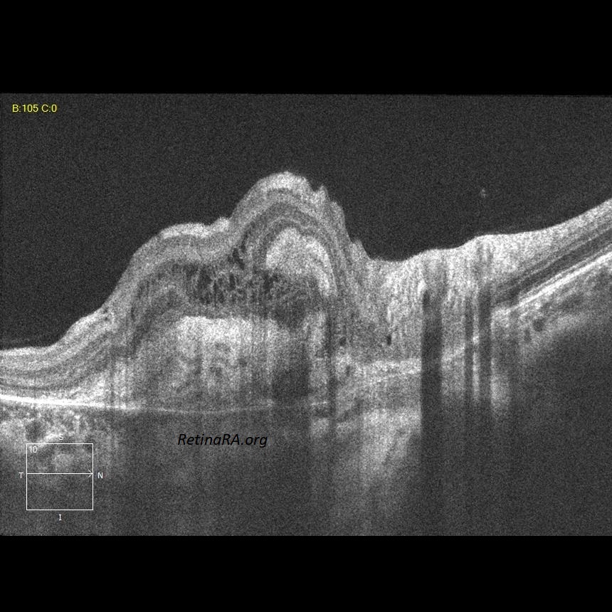


A 26-year-old female patient who complained of decreased visual acuity on the right eye was diagnosed with choroidal neovascular membrane (CNV) secondary to Birdshot retinochoroidopathy. Color photographs show peripapillary CNV in the right eye and bilateral peripapillary birdshot lesions; cream-coloured, irregular or elongated choroidal lesions whose long axis radiates from the optic disc. Atrophic birdshot lesions appear as hypoautofluorescent spots on the autofluorescence image. OCT reveals the CNV as hyperreflective subretinal and intraretinal tissue.
Credit: M. Giray Ersoz, MD, FEBO
Biruni University School of Medicine, Department of Ophthalmology, Istanbul, Turkey
Instagram accounts: @retina.review and @retina.dr.girayersoz