A 54-year-old woman without any systemic disease applied to the outpatient clinic due to presbyopic complaints. Her BCVA was 20/32 for both eyes and IOPs were within normal limits. Anterior segment examination was also unremarkable.
Dilated fundus examination showed bilateral optic nerve pits in both eyes, in addition to peripapillary round-shaped atrophy in the left eye. Both optic nerve pits were located temporally and no associated maculopathy was observed for each eye.
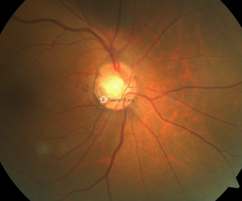
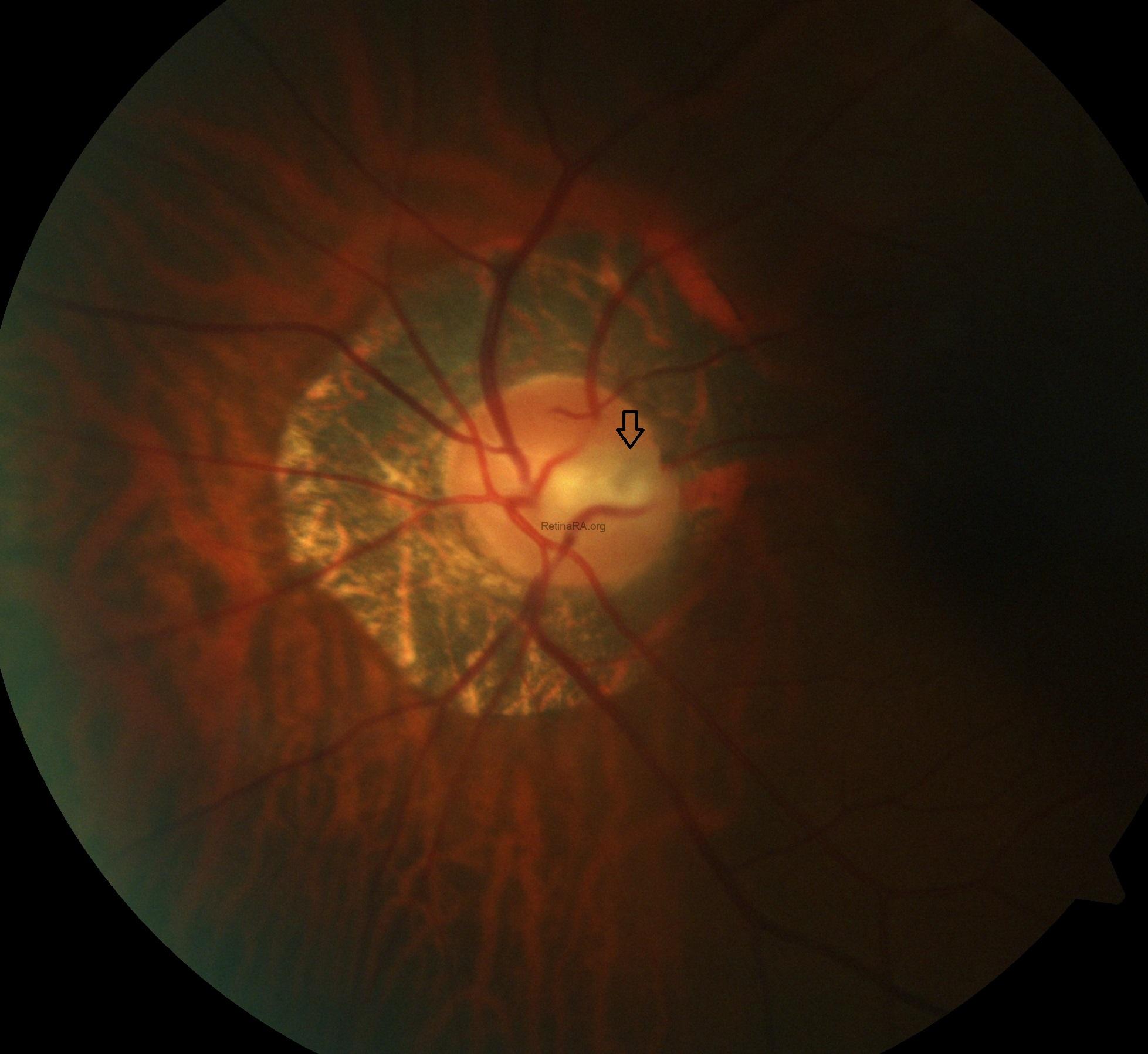
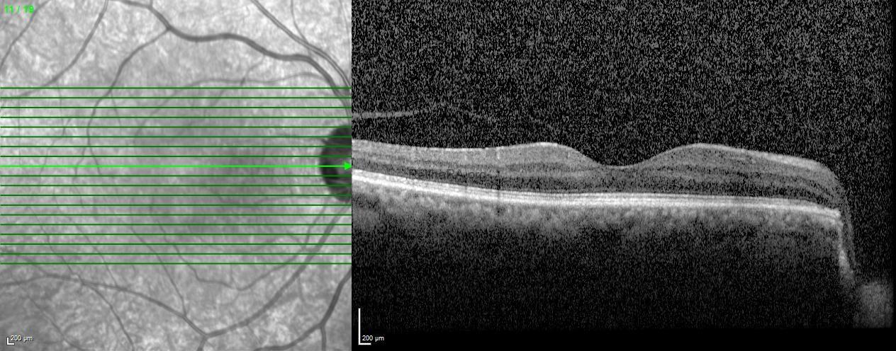
- Color fundus photography of the case with bilateral optic disc pit (right eye)
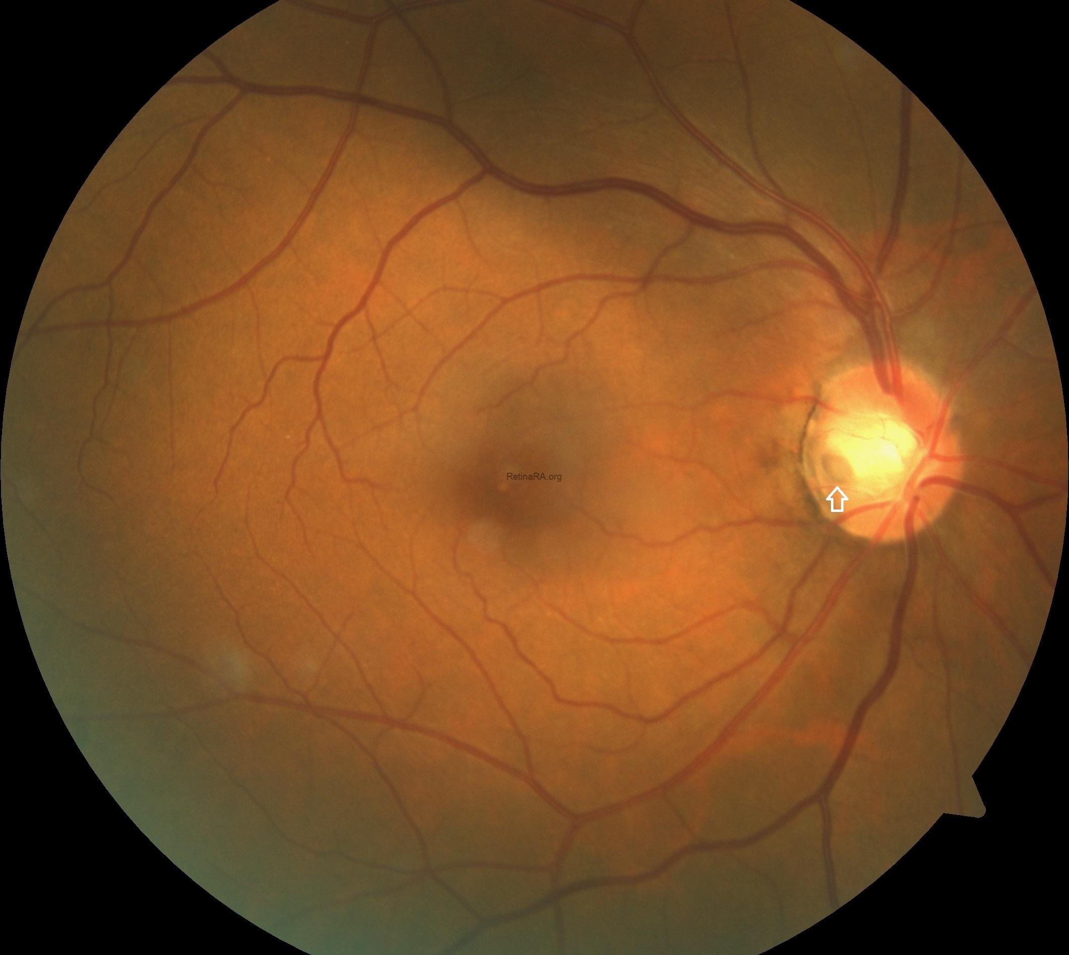
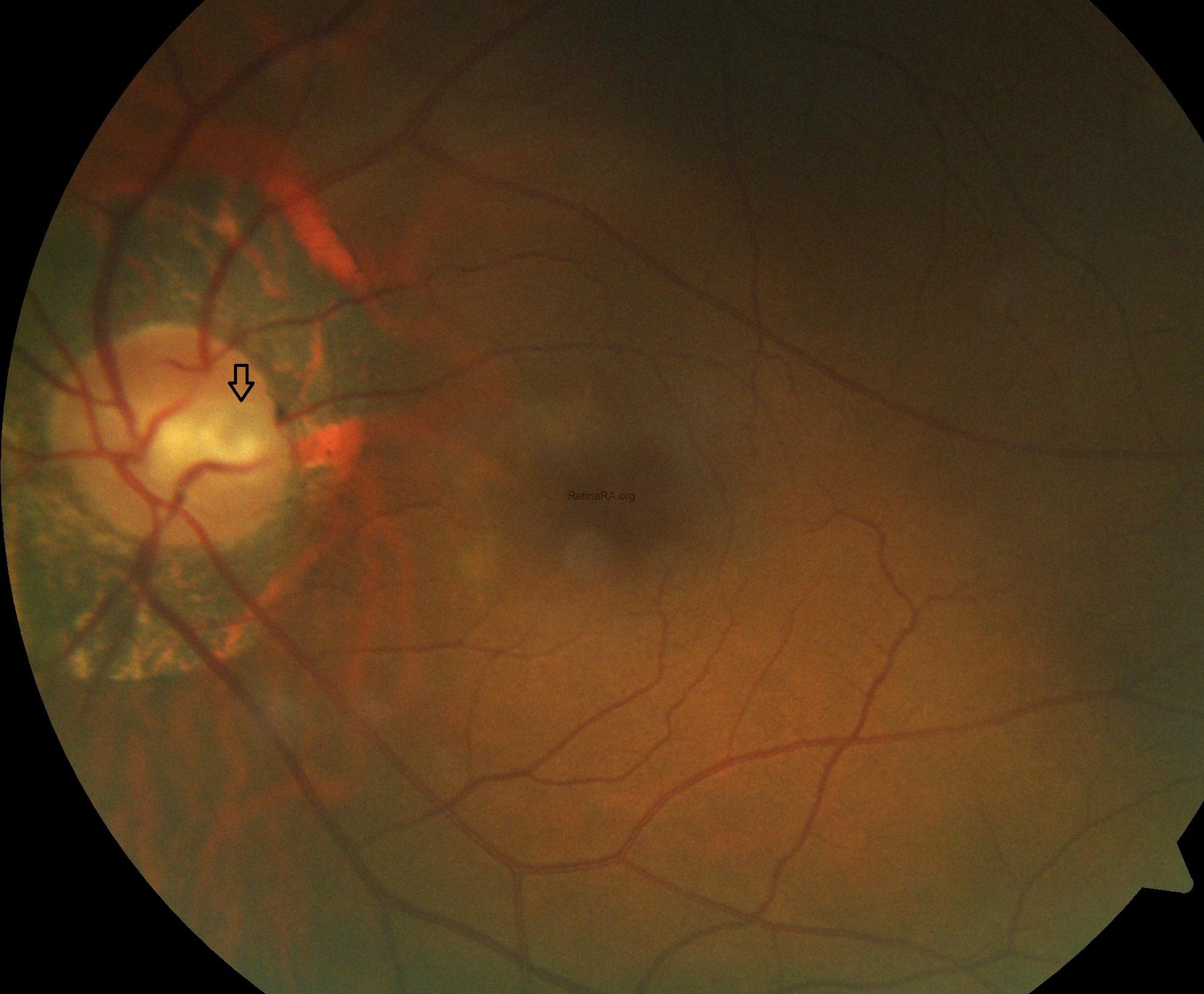
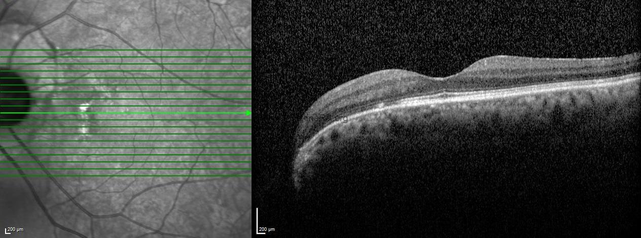
- Color fundus photography of the case with bilateral optic disc pit (left eye)
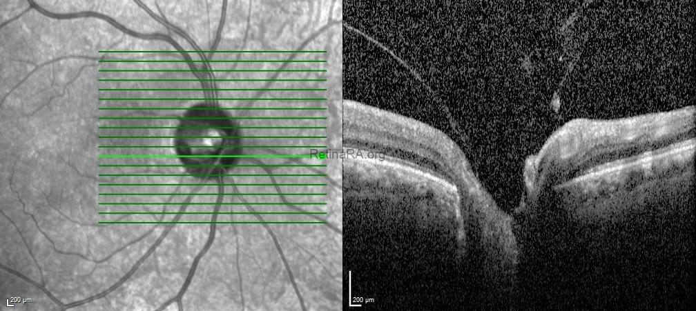
Optical coherence tomography scans confirmed the optic pits with attached glial tissue without any serous macular detachment and/or schisis-like inner retinal separation.
- OCT of the case with bilateral optic disc pit (right eye)
- OCT of the case with bilateral optic disc pit (left eye)
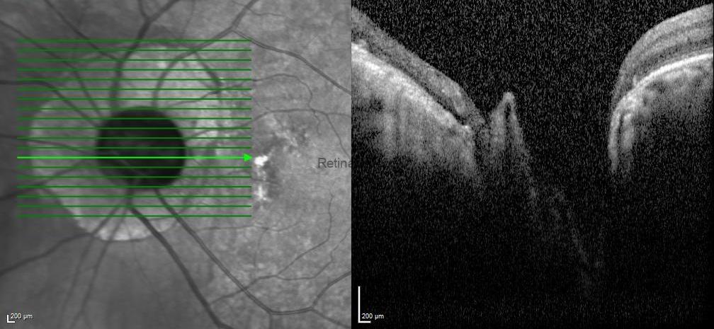
Optic disc pit is a rare congenital anomaly of the optic nerve head, often unilateral but occasionally seen bilaterally. While some patients remain asymptomatic, others may develop visual disturbances due to associated maculopathy, such as serous retinal detachment or schisis-like changes. Multimodal imaging, particularly OCT, is essential for detecting subtle structural alterations, monitoring progression, and guiding management. Although treatment strategies remain challenging and individualized, timely recognition is crucial for preserving visual function in these patients.
Credit: Kemal Tekin, M.D., from Ulucanlar Eye Training and Research Hospital
Instagram accounts: @retina.academy and @dr.kemaltekin