This was a 30 year-old male who was admitted with a history of low vision. The patient was healthy without any known systemic and ocular disease. The patient was a product of consanguinity marriage in which the parents of the patient were a second degree relative. The BCVAs were 20/70 and 20/30 for the right and left eyes, respectively and the intraocular pressures were within normal limits. The refractive errors of the patient were +1.50 diopters for the right and +1.75 for the left eye besides he had unremarkable anterior segment biomicroscopy findings.
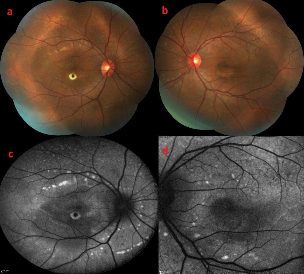
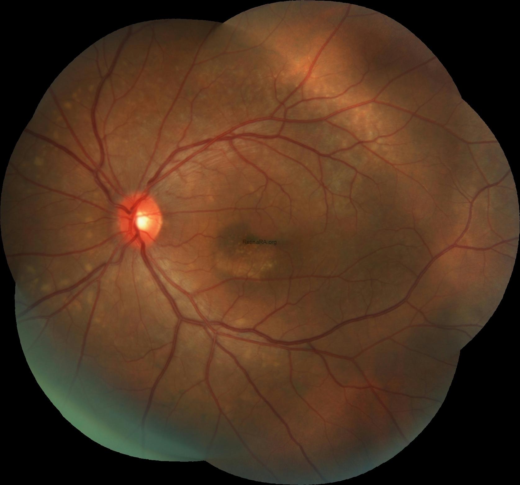
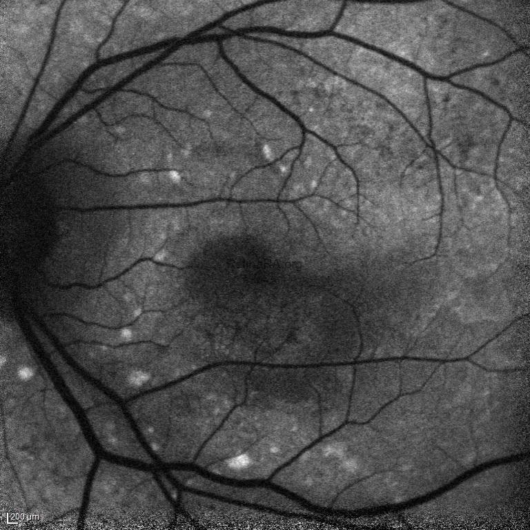
Dilated fundus examination demonstrated centrally hyperpigmented macular scar in the right eye and focal hypopigmented RPE atrophy at the inferior of fovea in the left eye in addition to bilateral multifocal subretinal white-yellow deposits in the macula and posterior pole of both eyes.
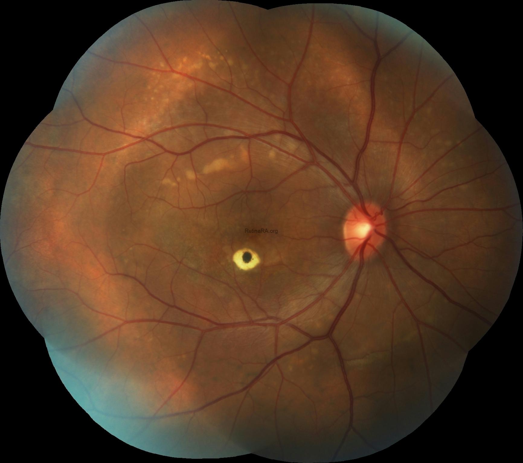
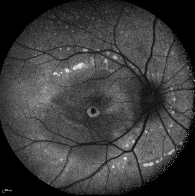
Fundus autofluorescence imaging exhibited that the central hyperpigmentation was hypo-autofluorescent while its surroundings were hyper- autofluorescent in the right foveal region with mixed or decreased autofluorescence at the inferior of fovea in the left eye as well as the deposits were brightly autofluorescent in both eyes.
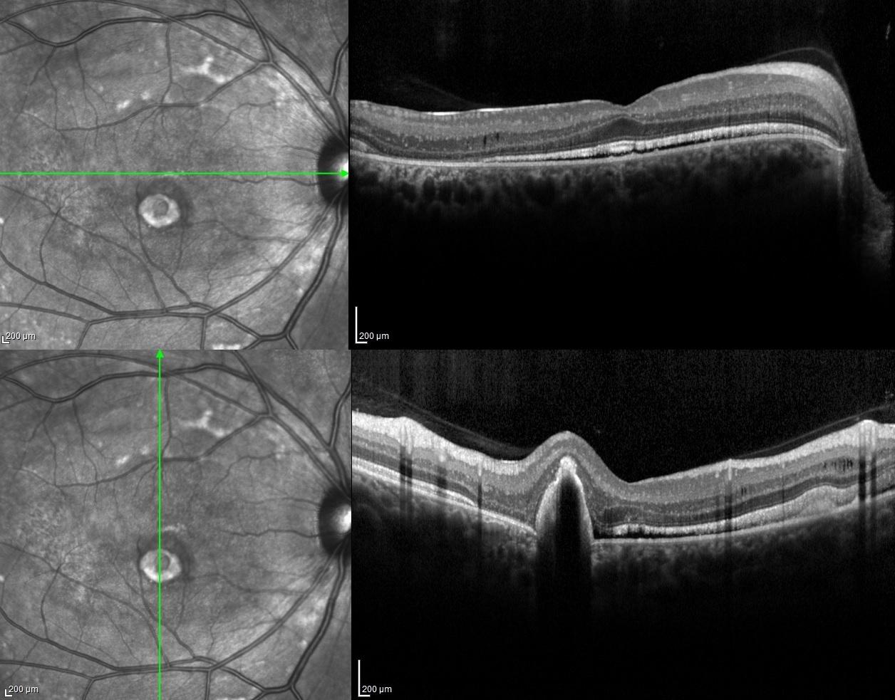
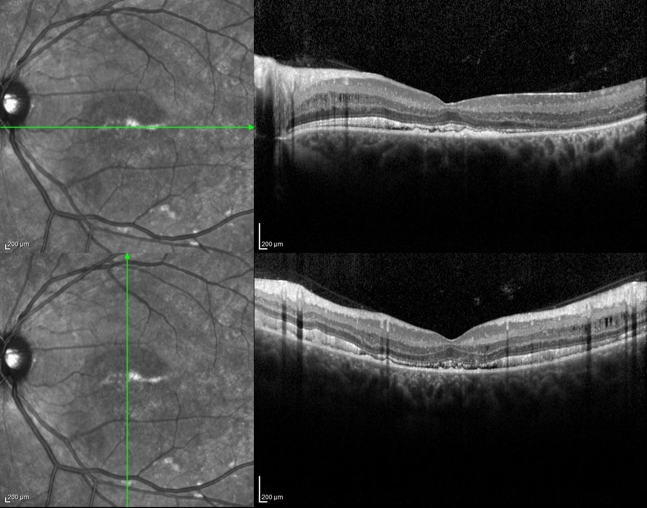
Spectral-domain optical coherence tomography of both eyes revealed minimally serous retinal detachment with dome shaped hyperreflective accumulation on RPE as well as retinoschisis in inner nuclear layers.
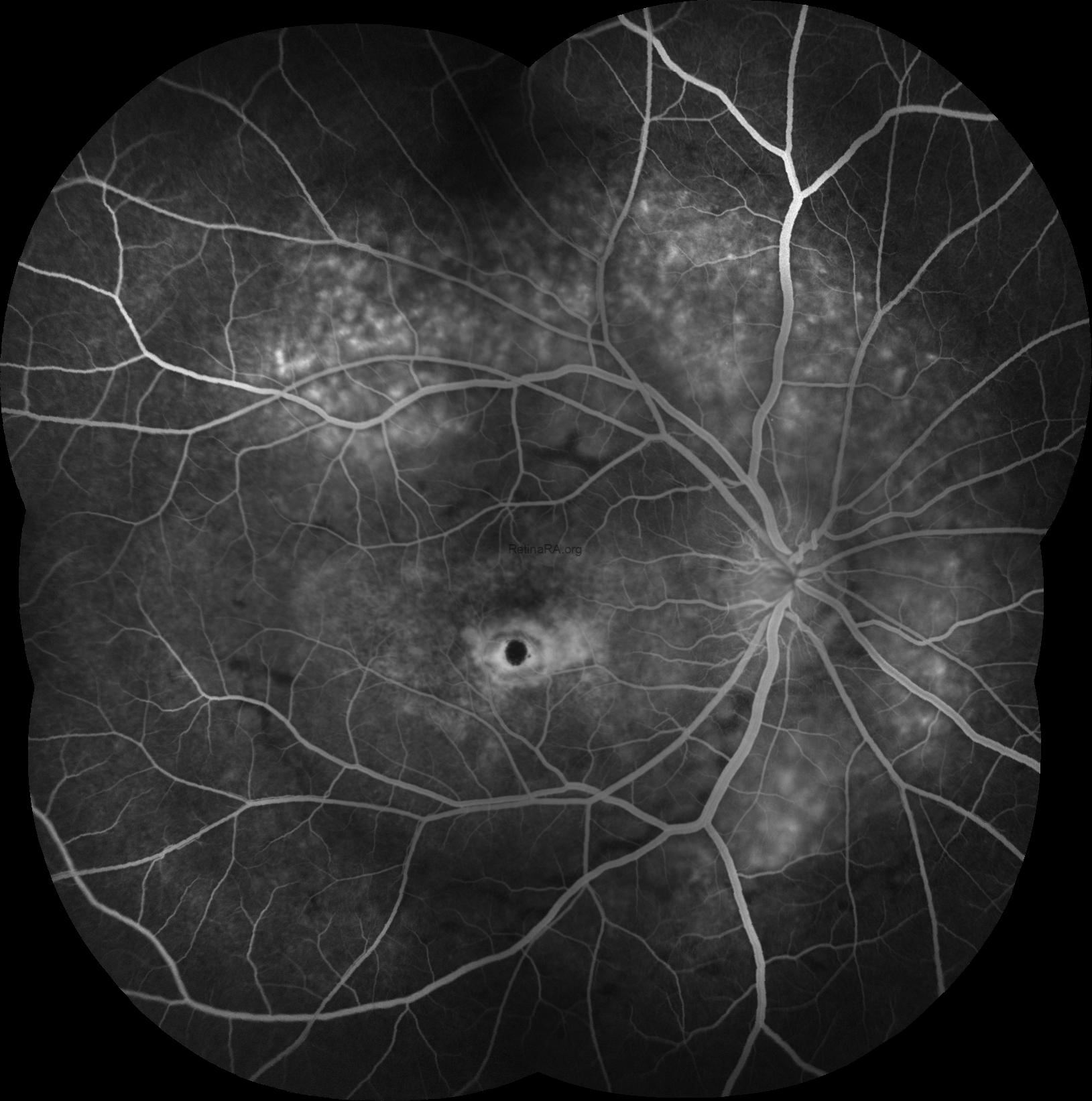
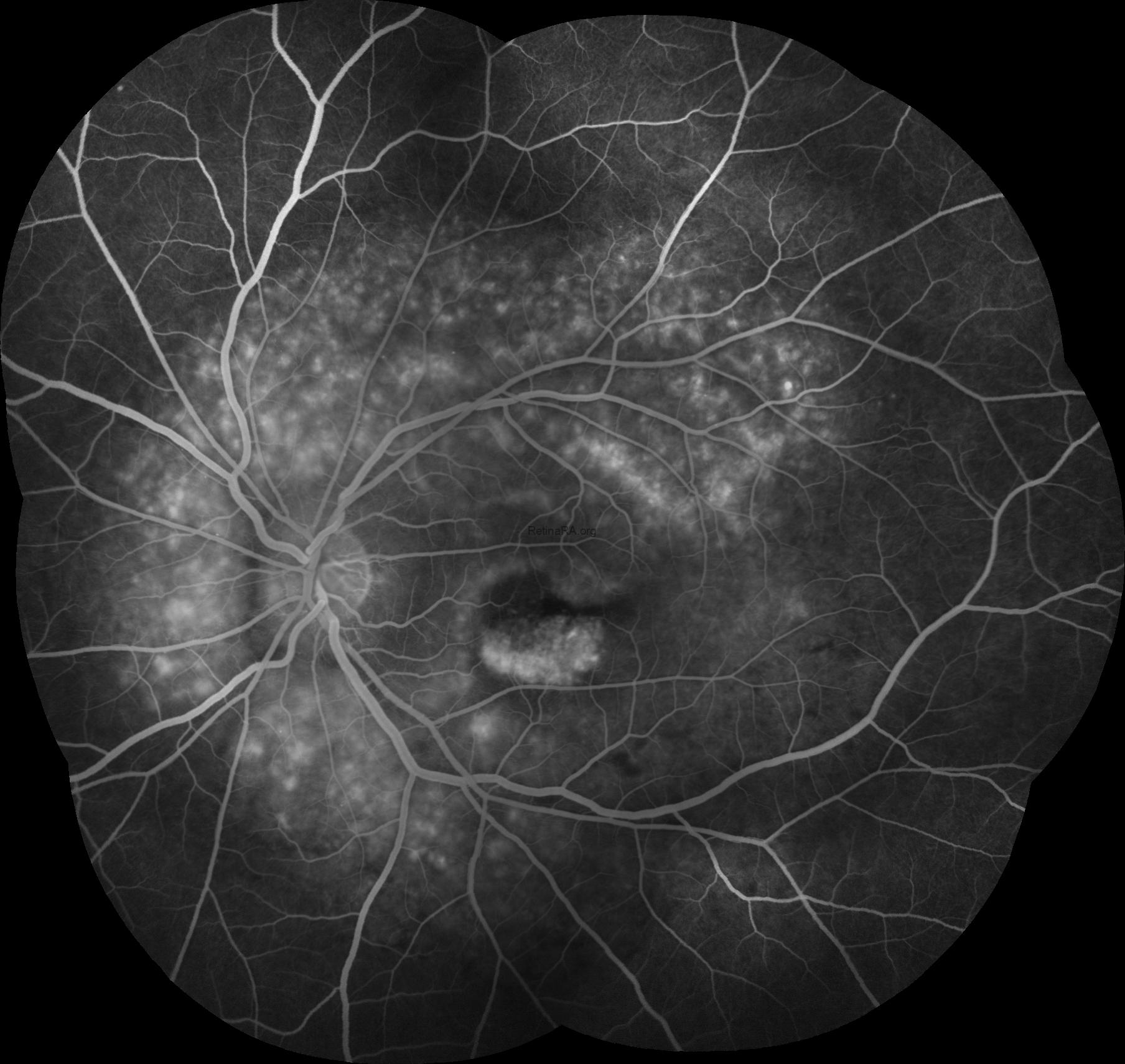
Fluorescein angiography imaging showed that the central hyperpigmentation was hypofluorescent due to blockage while its surroundings were window-defect hyperfluorescent in the right foveal region, with the window-defect hyperfluorescent at inferior of fovea in the left eye besides the staining of the subretinal deposits in both eyes.
Arden ratio was found as 1.18 for the right and 1.08 for the left eye in electrooculography.
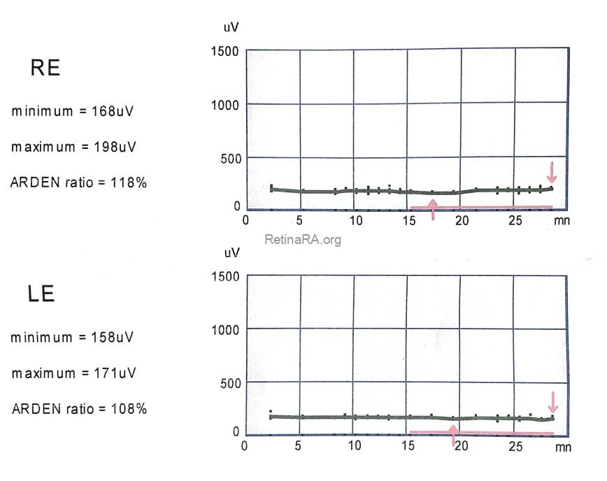
Autosomal recessive bestrophinopathy is caused by biallelic pathogenic variants in the BEST1 gene and has been first described in 2008 with findings consisting of central visual loss due to subretinal fluid or macular edema, characteristic retinopathy with the punctate flecks and vitelliform material deposition, an absent or severely reduced electrooculogram, abnormal or subnormal electroretinogram (ERG), and hyperopia with in some cases of shallow anterior chamber.
Credit: Kemal Tekin, M.D., from Ulucanlar Eye Training and Research Hospital
Instagram accounts: @retina.academy and @dr.kemaltekin
