34-year-old male was referred from the neurology department for the investigation of optic disc swelling etiology. Then patient had not any increased intracranial pressure symptoms such as headache and vomiting. The BCVAs were 5/10 for both eyes and intraocular pressures were within normal limits.
Fundus examinations of both eyes showed elevated optic disk with normal vascular features without any dilation, tortuosity and hemorrhages; and peripapillary atrophy with angioid streaks emanating from the optic nerve in addition to macular scar.


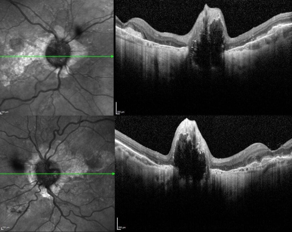
Spectral- domain optical coherence tomography scans passing through the optic nerve confirmed the elevated optic nerve and peripapillary chorioretinal atrophy.


Fundus autofluorescence imaging of both eyes demonstrated areas of increased autofluorescence at the nerve corresponding to the disc drusen and radial areas of hypo-autofluorescence corresponding to angioid streaks.


According to the multimodal imaging findings, the preliminary diagnosis was angioid streaks with optic disc drusen and the physical examination of the patient exhibited papular lesions and cutaneous laxity of the neck (“plucked chicken skin”) consistent with pseudoxanthoma elasticum.

Optic disk drusen are composed of small proteinaceous material which become calcified with advancing age. These deposits can be considered small tumors that develop within the optic nerve head, and might lead to an elevated disc (and thus this condition is sometimes referred to as pseudopapilledema). They can be associated with retinitis pigmentosa, angioid streaks, Usher syndrome, Noonan syndrome and Alagille syndrome.
ODD are more prevalent in Pseudoxanthoma elasticum (PXE) patients compared to populations with other ocular conditions. The exact etiology of this remains unknown. The hallmark pathological feature in PXE is calcification of Bruch membrane, starting around the optic nerve head and extending toward the periphery.
Credit: Kemal Tekin, M.D., from Ulucanlar Eye Training and Research Hospital
Instagram accounts: @retina.academy and @dr.kemaltekin
