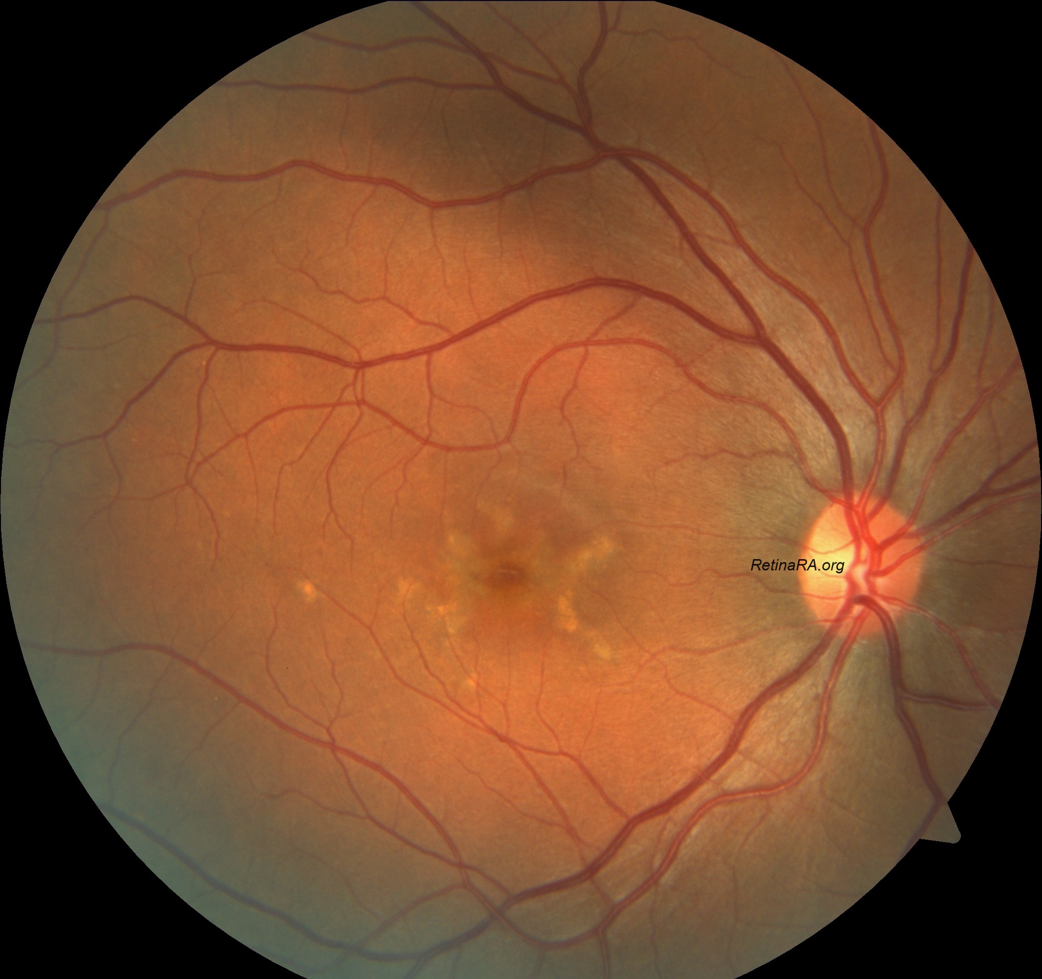
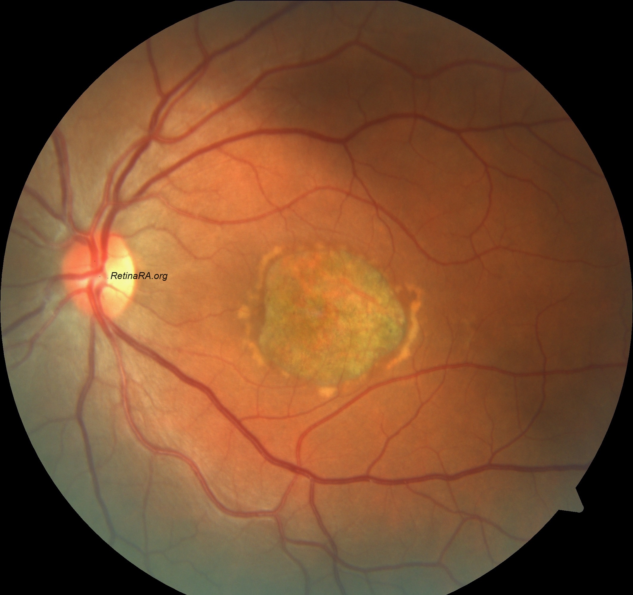
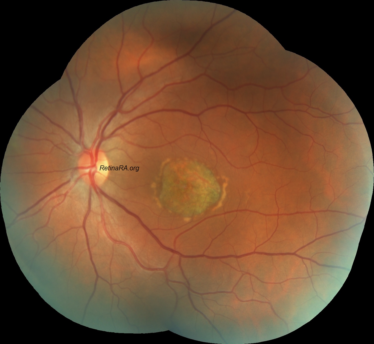
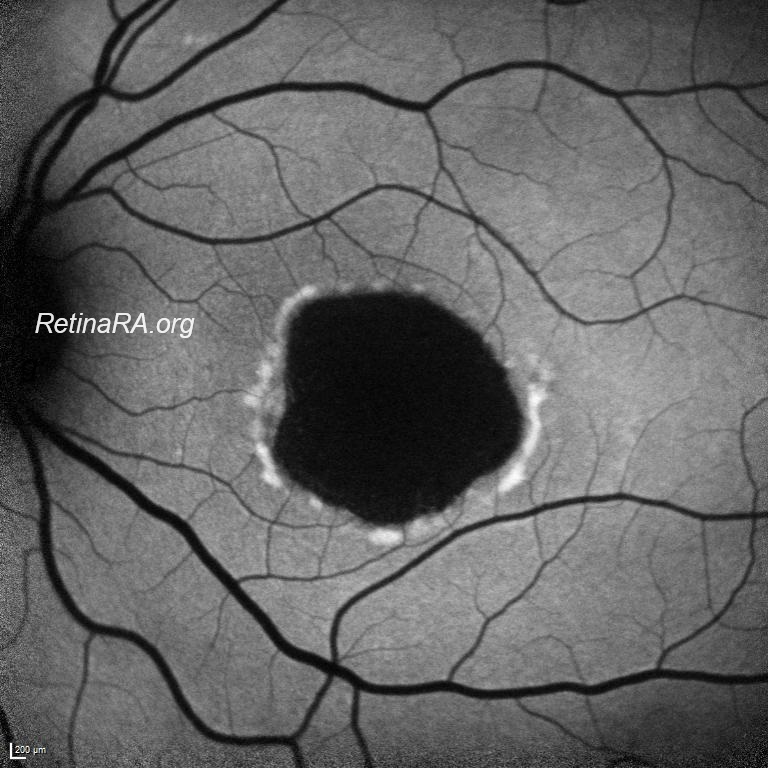
34-year old female presented with decreased vision in her both eyes. The BCVAs were 4/10 for the right eye and 1/10 for the left eye. Anterior segment examinations were unremarkable for both eyes. Colored fundus images showed abnormal foveal reflex with parafoveal flecks in the right eye and central sharply circumscribed chorioretinal atrophy in the macula with paramacular flecks in the left eye. Fundus autofluorescence imaging revealed hyper-autoflouresce of the flecks with hypo-autofluorescence of foveal atrophy. Optical coherence tomography exhibited atrophy of outer retinal layers in central macular areas in both eyes which was more prominent in the left eye. With these findings, ABCA4 retinopathy was preliminary diagnosis and confirmed with genetic analysis.
Stargardt disease and ABCA4-retinopathies are caused by pathogenic variants in the ABCA4 gene inherited in an autosomal recessive manner. Diagnosis of ABCA4-retinopathy is complex due to its phenotypic variability and the presence of other inherited retinal dystrophy phenocopies.
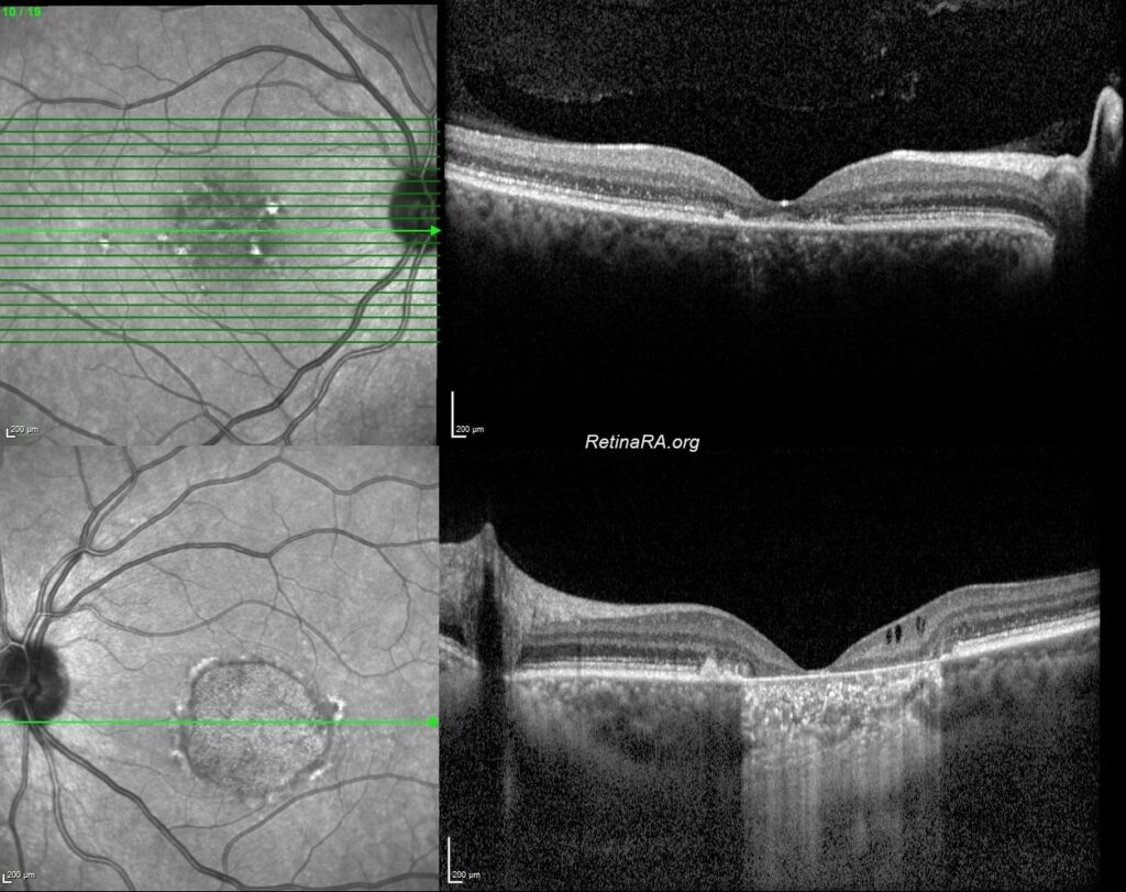
In patients with ABCA4-associated retinopathy, the characteristic clinical symptoms of the disease such as macular affection, fundus flecks, and peripapillary sparing can be observed. The fundal features are highly variable but patients usually have symmetrical ocular findings. The severity of these clinical symptoms can vary depending on the stage of the disease. ABCA4-retinopathy typically begins as a maculopathy which progresses to an enlarging lesion of outer retinal, RPE and choriocapillaris atrophy. This is usually accompanied by characteristic yellow flecks at the RPE level that are situated predominantly in the macula with variable peripheral distribution. SD-OCT reveals loss of normal architecture that begins at the central macula with relative preservation of the peripheral macula in the first instance and reduced central autofluorescence surrounded by an increased signal or a bull’s-eye maculopathy-like appearance on FAF.
Credit: Kemal Tekin, M.D., from Ulucanlar Eye Training and Research Hospital
Instagram accounts: @retina.academy and @dr.kemaltekin
Colored fundus image of the right eye of a patient with ABCA4-retinopathy.
Colored fundus image of the left eye of a patient with ABCA4-retinopathy.
Composed fundus image of the right eye of a patient with ABCA4-retinopathy.

Composed fundus image of the left eye of a patient with ABCA4-retinopathy.
Macular fundus autoflurescence of the of the right eye of a patient with ABCA4-retinopathy.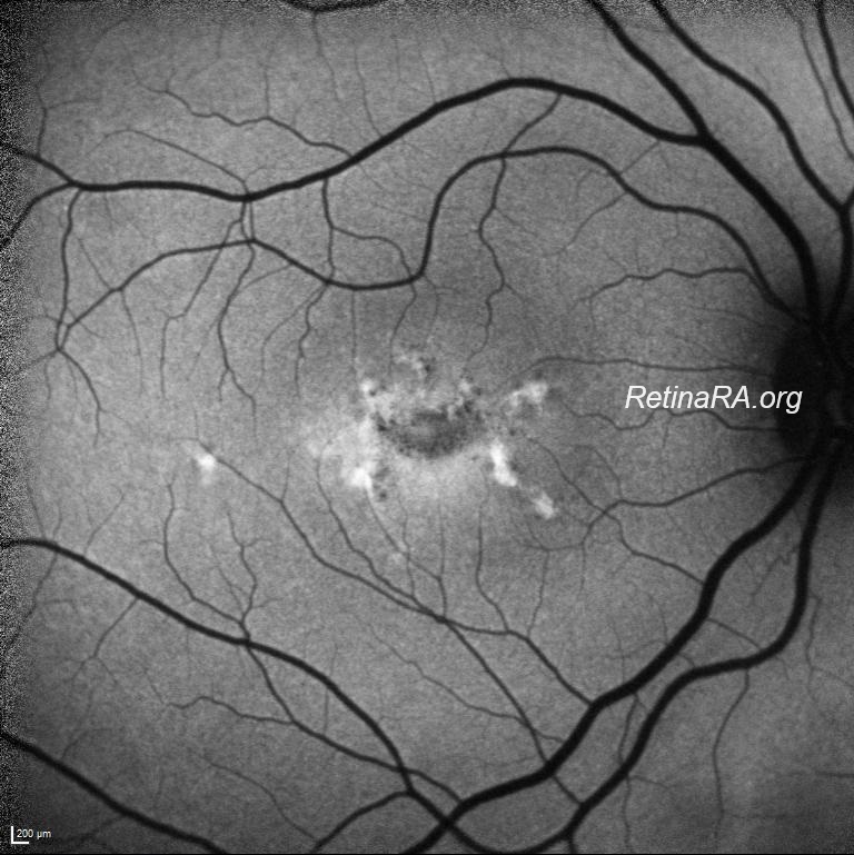
Fundus autoflurescence of the of the right eye of a patient with ABCA4-retinopathy.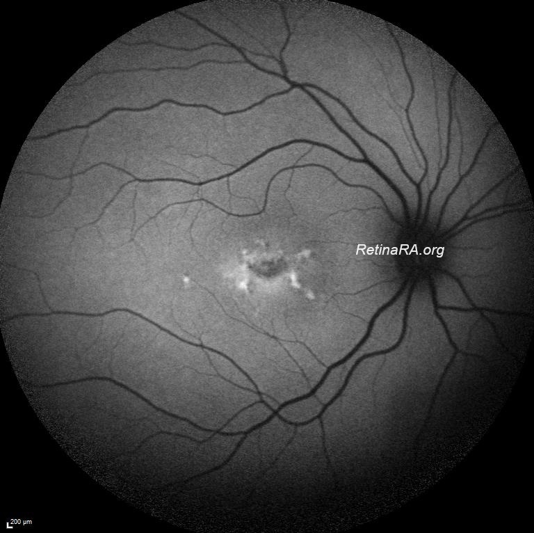
Macular fundus autoflurescence of the of the left eye of a patient with ABCA4-retinopathy.
Fundus autoflurescence of the of the right eye of a patient with ABCA4-retinopathy.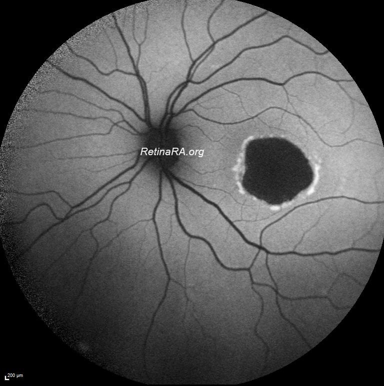
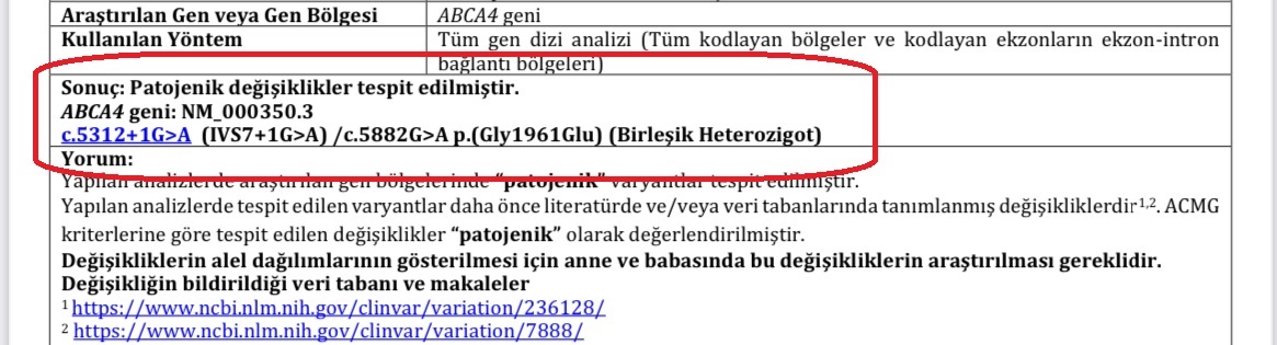
Genetic analysis of the patient.