
A 17-year-old male with a history of vitreoretinal surgery presented to the retina department for evaluation. Best-corrected visual acuity (BCVA) was 20/100 in the right eye and 20/60 in the left eye. Intraocular pressures and anterior segment examinations were unremarkable.
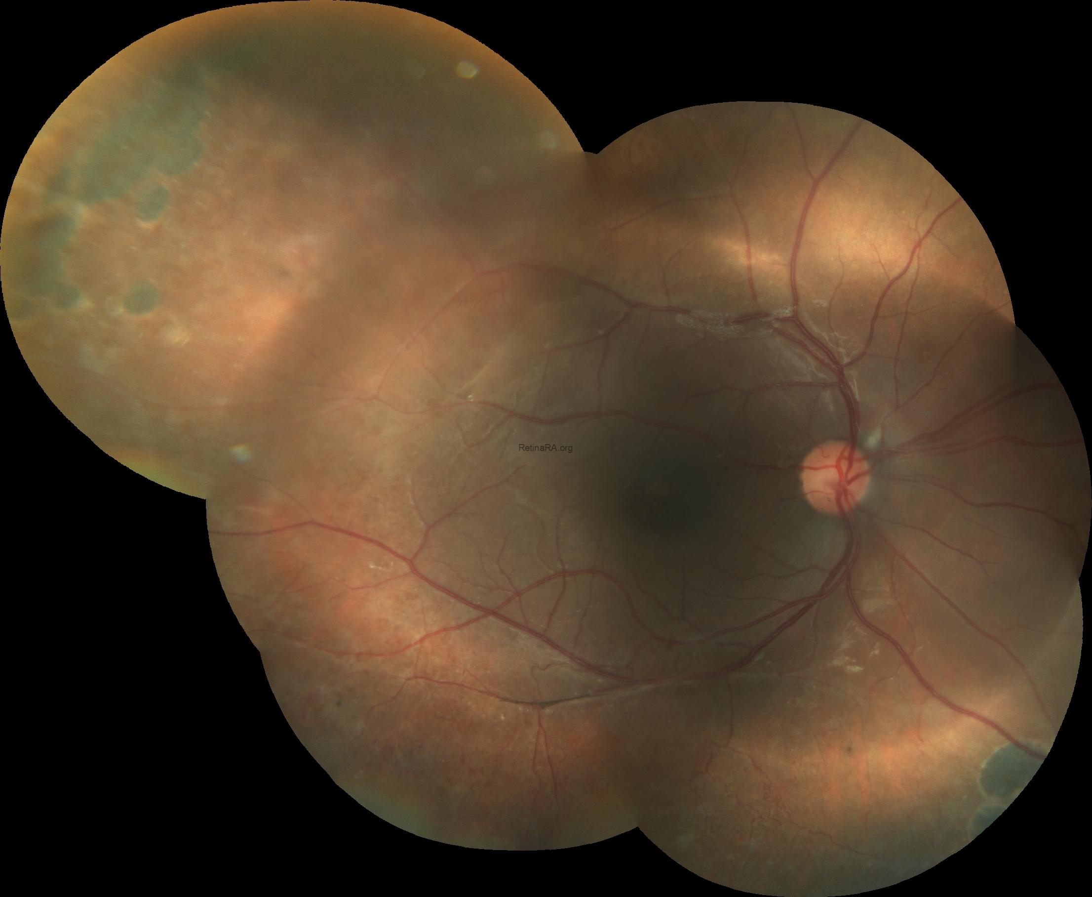
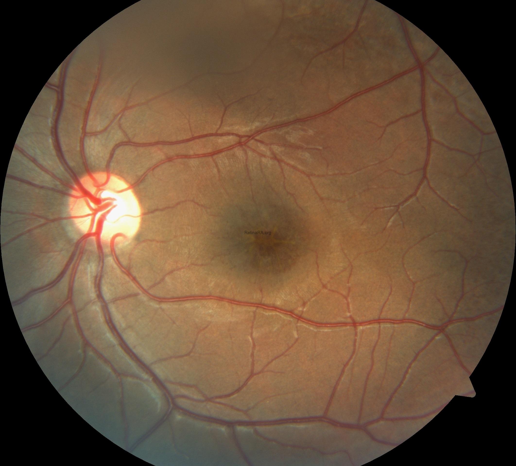
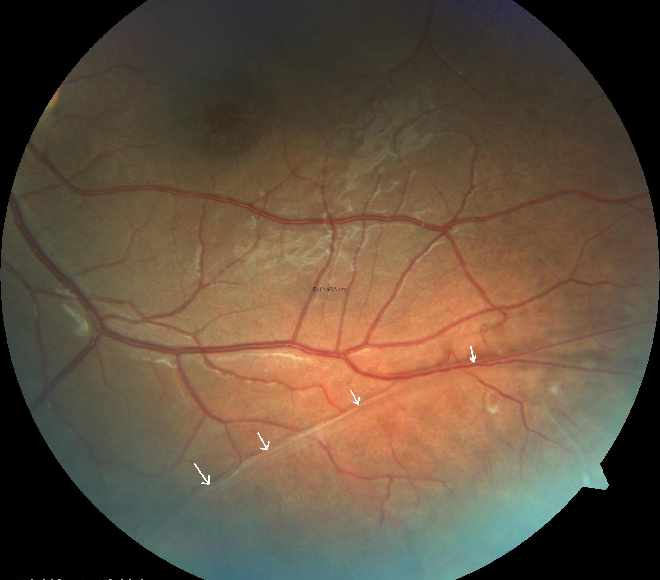
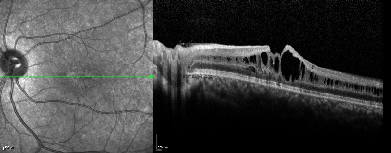
Fundus imaging revealed an abnormal foveal reflex with cystic lesions in both maculae. The right eye showed peripheral old laser scars and dome-shaped schisis, while the left eye demonstrated foveal schisis with a characteristic spoke-wheel pattern radiating from the fovea, along with an inferotemporal peripheral schisis.
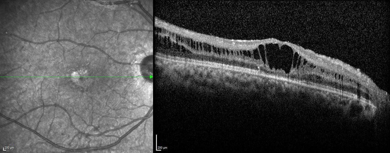
Optical coherence tomography (OCT) revealed diffuse cystoid spaces extending toward the optic nerve, causing splitting within the inner nuclear layer.
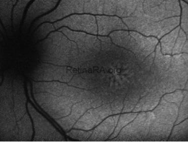
Fundus autofluorescence of the left eye showed alternating hyper- and hypoautofluorescent areas corresponding to the spoke-wheel pattern.
Based on these multimodal imaging findings, a preliminary diagnosis of X-linked retinoschisis was made, which was later confirmed by genetic testing revealing an RS1 gene mutation.
X-linked retinoschisis is a hereditary retinal disorder caused by mutations in the RS1 gene, leading to splitting of the retinal layers, primarily at the macula. It predominantly affects young males and is characterized by reduced central vision, cystic macular changes, and spoke-wheel patterns on OCT or fundus examination. Although there is no definitive cure, regular follow-up and management of complications such as vitreous hemorrhage or retinal detachment are crucial for preserving vision.
Credit: Kemal Tekin, M.D., from Ulucanlar Eye Training and Research Hospital
Instagram accounts: @retina.academy and @dr.kemaltekin